
BioCentral IBBioCentral IB
IB Biology · Theme B · B1.2 · SL and HL

Proteins

How a chain of amino acids folds into a working protein. Part one is for SL and HL; part two is HL only.
Guiding questions

What is the relationship between amino acid sequence and the diversity in form and function of proteins?

How are protein molecules affected by their chemical and physical environments?

Part one

Amino acids and polypeptides

B1.2.1 – B1.2.5
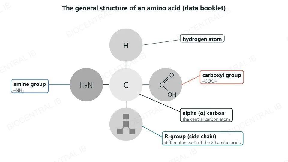
Data-booklet diagram of the general structure of an amino acid: a central alpha carbon joined to a hydrogen atom, an amine group (H2N), a carboxyl group (COOH) and an R-group, which differs in each of the 20 amino acids
B1.2.1

The structure of an amino acid

Every amino acid has the same general structure, shown in the data booklet.
  • A central carbon atom, the alpha (α) carbon, is bonded to four things: an amine group (–NH₂), a carboxyl group (–COOH), a hydrogen atom and an R-group.
  • The amine group, carboxyl group and hydrogen are the same in every amino acid.
  • Only the R-group (side chain) differs, giving each of the 20 amino acids in the genetic code its own properties.
SkillYou need to recognise and interpret this structure, not draw it from memory.
B1.2.2

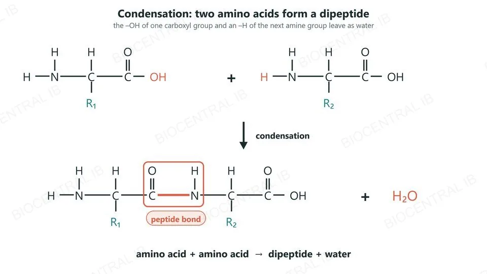
Peptide bonds: condensation

Amino acids join by condensation reactions, forming dipeptides and then longer chains.
  • The –OH of the carboxyl group of one amino acid and an –H of the amine group of the next leave as water.
  • A peptide bond forms between the carbon of the carboxyl group and the nitrogen of the amine group.
  • Word equation: amino acid + amino acid → dipeptide + water. Adding more amino acids the same way makes a polypeptide.
Try itBuild two amino acids with molecular models, remove a water molecule, then draw the dipeptide you have made.
Two amino acids with R-groups R1 and R2 join by condensation. The OH of the first carboxyl group and an H of the second amine group leave as water, forming a peptide bond between C and N. Word equation: amino acid plus amino acid gives dipeptide plus water
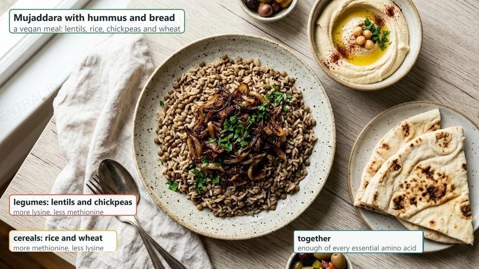
A vegan meal of mujaddara (lentils and rice) with hummus and bread. Legumes such as lentils and chickpeas have more lysine but less methionine; cereals such as rice and wheat have more methionine but less lysine; together they supply enough of every essential amino acid
B1.2.3

Essential amino acids

Essential amino acids cannot be synthesized by the body and must be obtained from food.
  • Non-essential amino acids can be made in the body from other amino acids.
  • Proteins in animal foods contain all the essential amino acids; many plant foods are low in one or more of them.
  • A vegan diet needs attention: combining legumes (such as lentils and chickpeas) with cereals (such as rice and wheat) supplies every essential amino acid.
ExampleMujaddara, lentils with rice, eaten with hummus and bread, is a vegan meal with a complete set of essential amino acids.
B1.2.4

An infinite variety of peptide chains

Twenty amino acids are coded for in the genetic code, and they can be joined in any order.
  • A chain can have any number of amino acids, from a few to thousands.
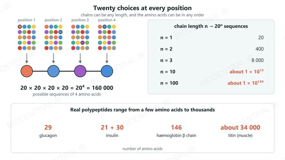
  • At each position there are 20 choices, so a chain of n amino acids could have 20ⁿ different sequences: about 1 × 10¹³ for just 10 amino acids.
  • Examples of polypeptides: glucagon (29 amino acids), the two chains of insulin (21 and 30) and titin in muscle (about 34 000).
Why it mattersThis almost unlimited variety is what lets proteins do so many different jobs.
Four positions in a chain, each with 20 possible amino acids, giving 20 to the power 4, or 160 000, sequences. A table: 20 to the power n is 20 for n = 1, 400 for 2, 8000 for 3, about 1 × 10 to the 13 for 10 and about 1 × 10 to the 130 for 100. Real polypeptides: glucagon 29 amino acids, insulin 21 + 30, the haemoglobin beta chain 146, titin about 34 000
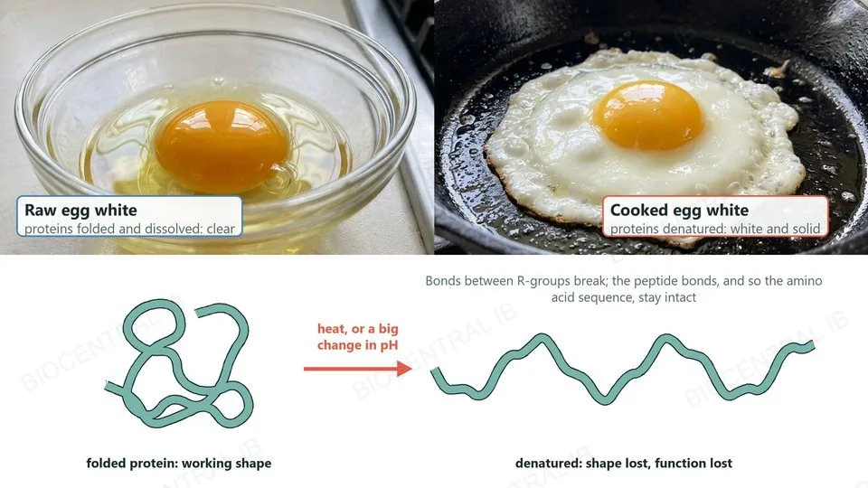
A raw egg white, clear because its proteins are folded and dissolved, beside a cooked egg white, white and solid because its proteins are denatured. Below, a folded protein chain unfolds when heated or when the pH changes; the bonds between R-groups break but the peptide bonds stay intact
B1.2.5

Heat, pH and denaturation

Denaturation is a change in the three-dimensional shape of a protein that stops it working.
  • High temperatures make the chain vibrate so much that the bonds between R-groups break and the protein unfolds.
  • A pH far from the normal value changes the charges on R-groups, breaking ionic bonds and hydrogen bonds.
  • The peptide bonds are not broken, so the sequence is unchanged, but the shape, and with it the function, is lost; often the change cannot be reversed.
ExampleCooking turns egg white from clear to white and solid as its proteins denature.
Quick check

A protein is heated strongly. It stops working, but its sequence of amino acids is unchanged. What has happened?

its peptide bonds have been broken by hydrolysis
bonds between R-groups broke and the chain unfolded
it has gained extra amino acids from the solution
its R-groups have been removed from the chain
Correct answer: bonds between R-groups broke. Heat breaks the hydrogen bonds, ionic bonds and other interactions that hold the folded shape. The peptide bonds survive, so the sequence is the same, but the protein is denatured.
Part two · HL

From R-groups to whole proteins

B1.2.6 – B1.2.12 · HL only
B1.2.6 · HL

The chemistry of R-groups

The chemical diversity of R-groups is the basis of the immense diversity in protein form and function.
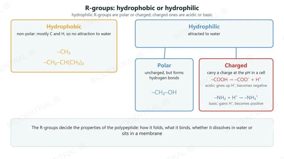
  • R-groups are either hydrophobic (non-polar) or hydrophilic.
  • Hydrophilic R-groups are polar or charged; charged R-groups are acidic or basic.
  • Amine and carboxyl groups in R-groups become charged by gaining or losing hydrogen ions: –NH₂ gains H⁺ to become –NH₃⁺, and –COOH loses H⁺ to become –COO⁻.
Why it mattersThe R-groups decide the properties of the polypeptide: how it folds, what it binds and where it can be.
R-groups are hydrophobic (non-polar, such as –CH3) or hydrophilic. Hydrophilic R-groups are polar (such as –CH2–OH) or charged. Charged R-groups are acidic (–COOH loses H+ to become –COO–) or basic (–NH2 gains H+ to become –NH3+)
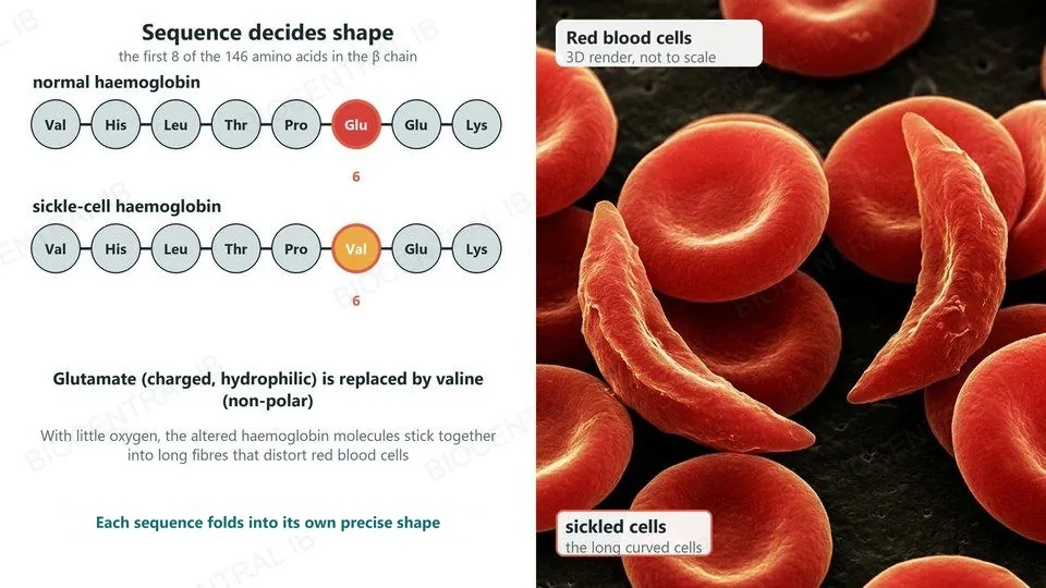
The first eight amino acids of the haemoglobin beta chain: Val His Leu Thr Pro Glu Glu Lys in normal haemoglobin and Val His Leu Thr Pro Val Glu Lys in sickle-cell haemoglobin, with position 6 highlighted. Beside it, a 3D render of red blood cells, with sickled cells among the normal discs
B1.2.7 · HL

Primary structure decides the shape

The primary structure, the sequence of amino acids, determines the three-dimensional shape of a protein.
  • The precise position of each amino acid decides which R-groups meet as the chain folds, and so which bonds form.
  • So proteins have precise, predictable and repeatable shapes: under the same conditions, copies with the same sequence normally fold the same way.
  • Changing one amino acid can change the shape: in sickle-cell haemoglobin, valine replaces glutamate at position 6 of the β chain, and the molecules stick together in fibres when oxygen is low.
LinkThe sequence itself is set by the gene (D1.2), so a mutation can alter the protein (D1.3).
B1.2.8 · HL

Secondary structure: the α-helix

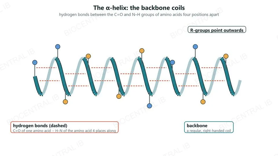
Secondary structure is the regular coiling or pleating of the backbone, held by hydrogen bonds in regular positions.
  • In an α-helix the chain coils into a regular helix.
  • Each C=O group in the backbone forms a hydrogen bond with the N–H group of the amino acid four places further along.
  • The R-groups point outwards from the helix, so they do not take part in these hydrogen bonds.
RememberSecondary structure uses the backbone; tertiary structure uses the R-groups.
An alpha helix: the backbone coils into a regular right-handed helix, hydrogen bonds (dashed) link the C=O of one amino acid to the N–H of the amino acid four places along, and R-groups point outwards
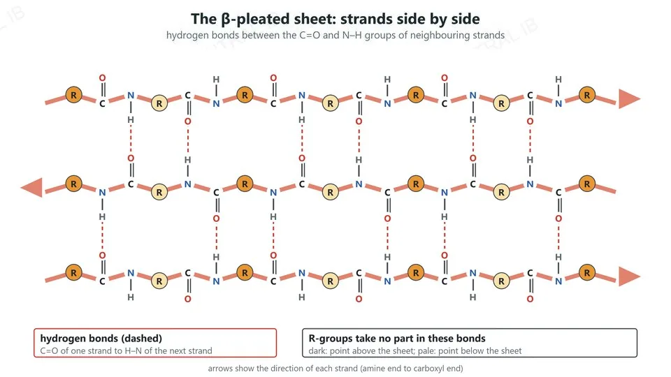
A beta-pleated sheet of three pleated strands side by side, the middle one running the opposite way, with hydrogen bonds between the C=O and N–H groups of neighbouring strands and R-groups, clear of those bonds, pointing above and below the sheet
B1.2.8 · HL

Secondary structure: the β-pleated sheet

In a β-pleated sheet, sections of the chain lie side by side and are linked by hydrogen bonds.
  • Each strand is pleated, folded in a zig-zag like a paper fan.
  • Hydrogen bonds form between C=O groups of one strand and N–H groups of the next, at regular positions.
  • The R-groups point alternately above and below the sheet.
Why it mattersMany hydrogen bonds together make helices and sheets stable, even though each bond is weak.
B1.2.9 · HL

Tertiary structure: hydrogen and ionic bonds

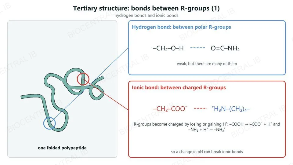
Tertiary structure is the overall three-dimensional folding of a polypeptide, held by bonds between R-groups.
  • Hydrogen bonds form between polar R-groups.
  • Ionic bonds form between R-groups with opposite charges, such as –NH₃⁺ and –COO⁻.
  • R-groups become charged by binding or releasing hydrogen ions, so a change in pH can break ionic bonds.
LinkThis is why pH can denature a protein (B1.2.5).
A folded polypeptide with two marked sites: a hydrogen bond between polar R-groups, –CH2–OH and O=C–NH2, and an ionic bond between charged R-groups, –CH2–COO– and +H3N–(CH2)4–. R-groups become charged by losing or gaining H+, so pH affects ionic bonds
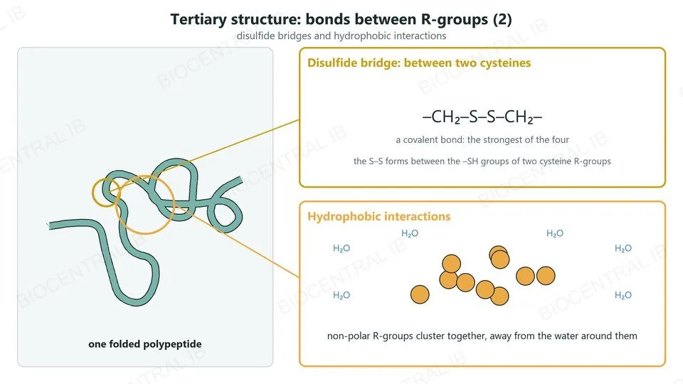
A folded polypeptide with two marked sites: a disulfide bridge, –CH2–S–S–CH2–, a covalent bond between two cysteine R-groups, and a cluster of non-polar R-groups held together by hydrophobic interactions away from water
B1.2.9 · HL

Tertiary structure: disulfide and hydrophobic

Two more kinds of bonding hold the folded chain in shape.
  • Disulfide bonds are covalent bonds between the sulfur atoms of two cysteine R-groups: the strongest of the four.
  • Hydrophobic interactions hold non-polar R-groups together, away from water.
  • Cysteine is the only amino acid you need to name: pairs of cysteines form disulfide bonds.
SummaryTertiary structure: hydrogen bonds, ionic bonds, disulfide bonds and hydrophobic interactions.
B1.2.10 · HL

Polar and non-polar amino acids

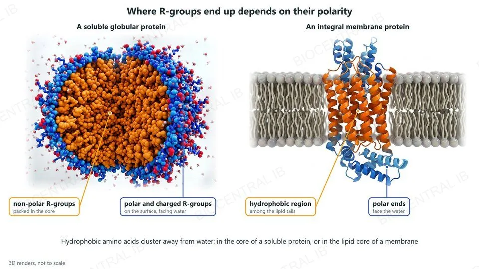
Where each amino acid ends up in a folded protein depends on whether its R-group is polar or non-polar.
  • In proteins that dissolve in water, hydrophobic amino acids cluster in the core of the globular protein.
  • Polar and charged amino acids sit on the surface, where they interact with water and keep the protein soluble.
  • Integral membrane proteins have regions of hydrophobic amino acids that embed them in the hydrophobic core of the membrane.
LinkB2.1: integral and peripheral proteins in membranes.
Two 3D renders. A soluble globular protein cut in half: non-polar R-groups packed in its core, polar and charged R-groups on the surface facing water. An integral membrane protein: a hydrophobic region among the lipid tails and polar ends facing the water on each side
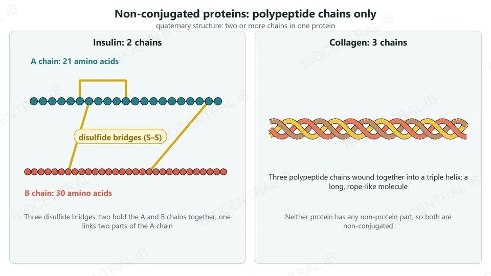
Insulin: an A chain of 21 amino acids and a B chain of 30 amino acids, held by disulfide bridges, two between the chains and one within the A chain. Collagen: three polypeptide chains wound into a triple helix. Neither has a non-protein part
B1.2.11 · HL

Quaternary structure: non-conjugated

Quaternary structure is the linking of two or more polypeptides into one protein.
  • Non-conjugated proteins are made only of polypeptides.
  • Insulin has two chains, A (21 amino acids) and B (30), held together by disulfide bridges.
  • Collagen has three polypeptide chains wound together into a triple helix.
ExamplesInsulin and collagen: quaternary structure without any non-protein part.
B1.2.11 · HL

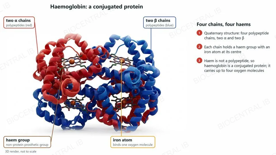
Quaternary structure: conjugated

A conjugated protein has one or more non-polypeptide parts, called prosthetic groups.
  • Haemoglobin has four polypeptide chains: two α chains and two β chains.
  • Each chain holds a haem group, a non-protein prosthetic group with an iron atom at its centre.
  • Each haem can bind one oxygen molecule, so one haemoglobin molecule can carry four.
ExampleHaemoglobin is the guide’s example of a conjugated protein.
A 3D render of haemoglobin: two alpha chains (red) and two beta chains (blue), each holding a haem group, a non-protein prosthetic group with an iron atom at its centre that binds one oxygen molecule. Haemoglobin is a conjugated protein
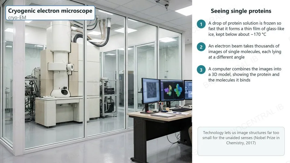
A cryogenic electron microscope in a laboratory, with a scientist at screens showing a micrograph and a 3D protein model. Steps: the protein solution is frozen into a thin film of glass-like ice; an electron beam images thousands of single molecules; a computer combines them into a 3D model
B1.2.11 · HL · Nature of science

Imaging proteins: cryo-EM

Technology allows imaging of structures that would be impossible to observe with the unaided senses.
  • In cryogenic electron microscopy (cryo-EM), proteins are frozen very fast in a thin layer of ice and imaged with electrons.
  • Thousands of images of single molecules, lying at different angles, are combined by computer into a 3D model.
  • Cryo-EM has shown single protein molecules and how they interact with other molecules; its developers won the 2017 Nobel Prize in Chemistry.
Nature of scienceNew instruments extend our senses and so open new areas of science.
B1.2.7 – B1.2.11 · HL

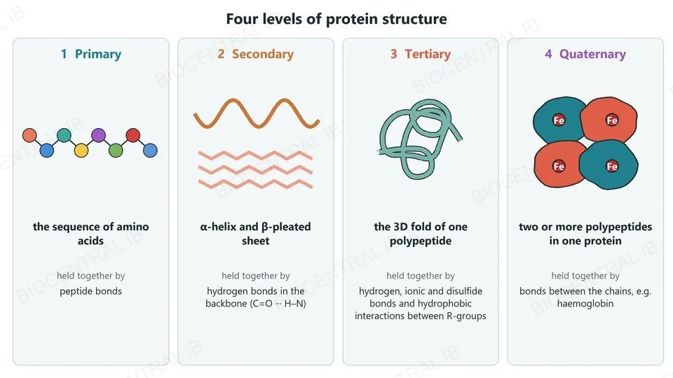
Four levels of protein structure

Each level builds on the one before, and each is held by its own kind of bonding.
  • Primary: the sequence of amino acids, joined by peptide bonds.
  • Secondary: α-helices and β-pleated sheets, held by hydrogen bonds in the backbone.
  • Tertiary: the 3D fold of one polypeptide, held by bonds between R-groups.
  • Quaternary: two or more polypeptides in one protein, such as haemoglobin.
Why it mattersThe sequence at level one decides every level above it.
Four levels of protein structure: primary, the sequence of amino acids held by peptide bonds; secondary, alpha helix and beta-pleated sheet held by backbone hydrogen bonds; tertiary, the 3D fold of one polypeptide held by bonds between R-groups; quaternary, two or more polypeptides in one protein, such as haemoglobin
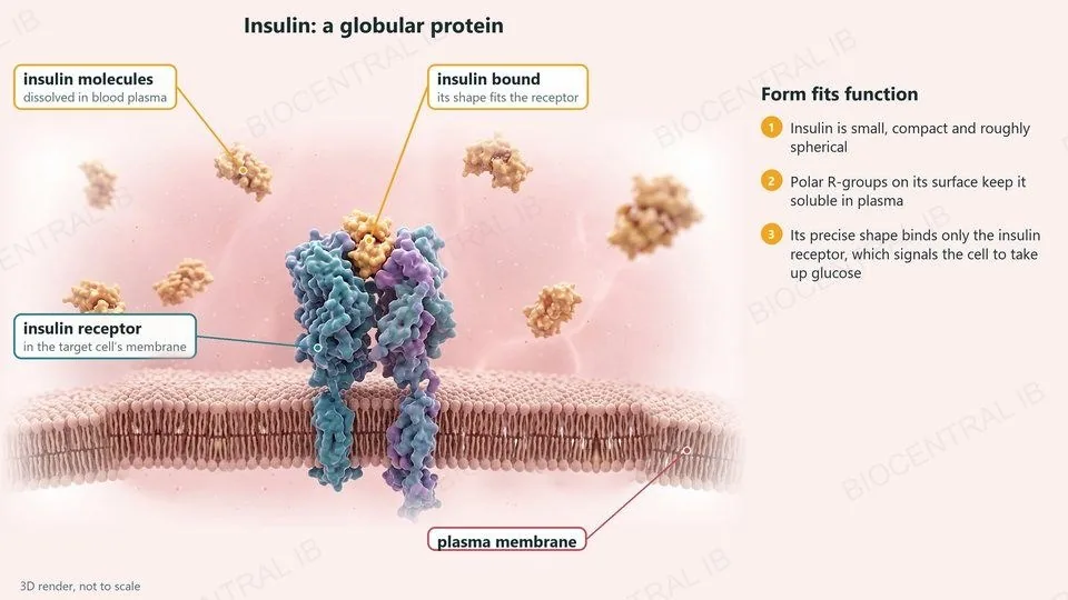
A 3D render of compact insulin molecules dissolved in blood plasma above a cell membrane; one insulin molecule is bound to the insulin receptor in the target cell’s membrane, because its precise shape fits the receptor. Polar R-groups on its surface keep it soluble
B1.2.12 · HL

Globular proteins: insulin

Globular proteins are compact and roughly spherical, and their shapes suit them to specific functions.
  • Insulin is a small globular hormone that travels in blood plasma.
  • Polar R-groups on its surface keep it soluble in the plasma.
  • Its precise shape fits only the insulin receptor on target cells, so it signals only those cells, telling them to take up glucose.
Form and functionCompact and soluble with a precise shape: ideal for a signal carried in the blood.
B1.2.12 · HL

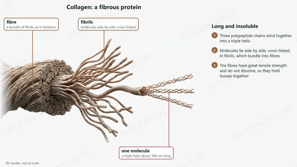
Fibrous proteins: collagen

Fibrous proteins are long and narrow, and their shapes suit them to structural roles.
  • Collagen molecules are triple helices about 300 nm long.
  • Molecules lie side by side, cross-linked, in fibrils, which bundle into fibres in tendons, skin and bone.
  • The fibres have great tensile strength and do not dissolve, so they hold tissues together.
CompareInsulin is compact and soluble; collagen is long and insoluble. Each shape suits its function.
A 3D render of collagen at three scales: a fibre, a bundle of fibrils as in tendons; fibrils, molecules side by side and cross-linked; and single molecules, each a triple helix about 300 nm long. The fibres are strong and insoluble
Quick check · HL

In a protein that dissolves in water, where are the non-polar R-groups usually found, and why?

on the surface, to attract water molecules
in the core, away from water
in disulfide bridges with cysteine
evenly spread through the whole protein
Correct answer: in the core. Non-polar R-groups are hydrophobic, so the chain folds with them clustered inside, held by hydrophobic interactions. Polar and charged R-groups on the surface face the water and keep the protein soluble.

Key vocabulary

Worth being able to define in a single sentence each

Amino acid
an α-carbon with an amine group, carboxyl group, H and R-group.
Peptide bond
the C–N bond formed when two amino acids condense.
Polypeptide
a chain of many amino acids joined by peptide bonds.
Essential amino acid
cannot be made by the body, so must come from food.
Denaturation
loss of a protein’s 3D shape, and so its function.

Key vocabulary · HL

Additional higher level terms

Primary structure
the sequence of amino acids in a polypeptide.
Secondary structure
α-helix or β-sheet, held by backbone hydrogen bonds.
Tertiary structure
the 3D fold of one polypeptide, held by R-group bonds.
Quaternary structure
two or more polypeptides linked in one protein.
Conjugated protein
includes a non-protein prosthetic group, e.g. haem.
Globular / fibrous
compact and soluble / long and insoluble.

Where this shows up again

A1.2 / D1.2 · Nucleic acids, protein synthesis
Ribosomes make proteins using the genetic code. How does the base sequence of DNA decide the primary structure of a protein?
D1.3 / D3.2 · Mutation, inheritance
Using sickle-cell disease, explain how a change in primary structure can alter quaternary structure and reduce a protein’s function.
C1.1 · Enzymes
How do the effects of temperature and pH on protein structure explain how enzyme activity changes?
B2.1 · Membranes
How do hydrophobic and hydrophilic amino acids let integral proteins sit in a membrane?

B1.2 Proteins — one-page recap

Screenshot this slide to revise from

Amino acids
  • α-carbon + amine group + carboxyl group + H + R-group.
  • Condensation forms peptide bonds: amino acid + amino acid → dipeptide + water.
Diet and variety
  • Essential amino acids must come from food; vegans combine legumes and cereals.
  • 20 amino acids in any order and number: 20ⁿ possible sequences.
Denaturation
  • Heat or pH breaks bonds between R-groups; the shape and function are lost.
  • Peptide bonds stay intact.
HL · R-groups and folding
  • Hydrophobic or hydrophilic (polar, or charged: acidic or basic).
  • Sequence decides shape; secondary structure: α-helix, β-sheet (backbone H-bonds).
HL · Tertiary
  • Hydrogen, ionic, disulfide bonds and hydrophobic interactions between R-groups.
  • Non-polar core in soluble proteins; hydrophobic belts in membrane proteins.
HL · Quaternary and shape
  • Insulin, collagen non-conjugated; haemoglobin conjugated (haem).
  • Globular insulin vs fibrous collagen: form suits function.
Next step

Now test yourself on B1.2

Reading the slides is half the job. The other half is answering questions the way the exam asks them.

36 quiz questions7 data questions5 exam questionsmarkschemes included

These study notes are free to read. Unlock every lesson with Pro — quizzes, data questions and exam papers. Two lessons are completely free to try: A1.1 Water and B1.1 Carbohydrates and lipids.

One-page summary, key terms and sample questions for B1.2 →

Sequence becomes shape, and shape becomes function.

Twenty amino acids, joined and folded, build every enzyme, hormone and fibre.
B1.2 Proteins · BioCentral IB
Use ↓ ↑ or click to navigate
01 / 20