BioCentral IBBioCentral IB
IB Biology · Theme D · D1.1

DNA replication

Before every cell division, an entire genome must be copied letter for letter. Helicase unwinds the double helix, DNA polymerase builds two identical daughter molecules strand by strand, and that same enzyme toolkit now powers PCR, DNA profiling, and forensic science far beyond the cell.
Guiding questions

How is new DNA produced?

How has knowledge of DNA replication enabled applications in biotechnology?

Part one

Copying the genome accurately

D1.1.1 – D1.1.3
D1.1.1

Every division needs an exact copy

DNA replication produces two DNA molecules with base sequences identical to the original — an exact copy, not merely a similar one.
  • Every time a cell divides by mitosis, each daughter cell needs its own complete, identical copy of the genome, so replication must be finished before division begins.
  • In multicellular organisms, this same copying process underlies growth (increasing cell number) and the ongoing replacement of damaged or worn-out tissue.
  • Reproduction depends on it too: an entire new organism arises from repeated rounds of replication and division, starting from one copied genome.
Why it mattersReplication copies; it does not mix alleles. New combinations of alleles come from meiosis and fertilization, and the rare copying errors that slip through become mutations (D1.3).
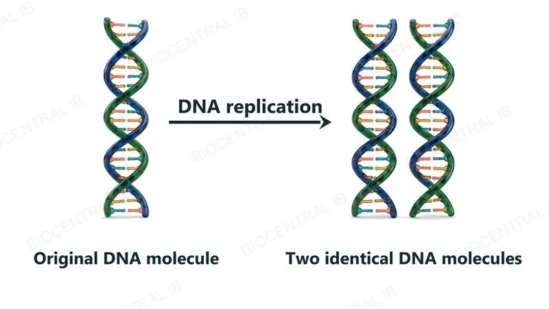
One DNA double helix on the left, labelled Original DNA molecule, and an arrow labelled DNA replication pointing to two identical DNA double helices on the right, labelled Two identical DNA molecules
D1.1.2

Half old, half new

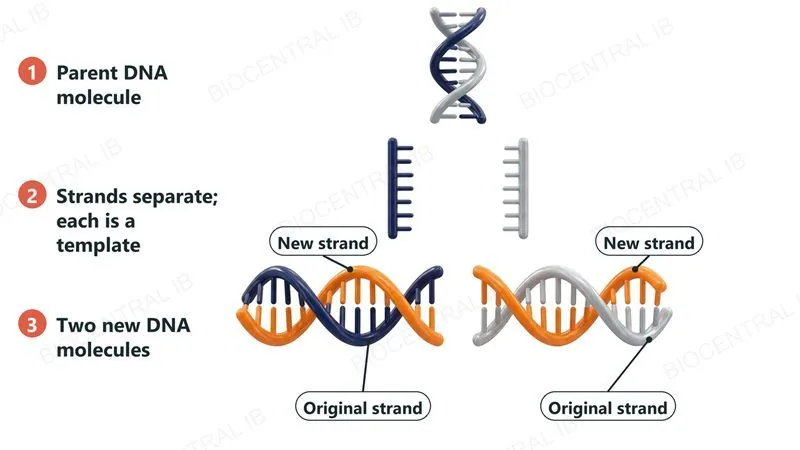
DNA replication is semi-conservative: each new DNA molecule keeps one original strand and pairs it with one newly built strand.
  • The two original strands separate, and each acts as a template; complementary base pairing (A with T, C with G) fixes exactly which new nucleotide is added opposite each template base.
  • Building each new strand by matching bases to an existing template, rather than assembling one from scratch, is what gives replication its high accuracy.
  • Meselson and Stahl's density-gradient experiment, tracking heavy ¹⁵N-labelled DNA across generations, provided the classic evidence for this semi-conservative model.
Why it mattersComplementary base pairing is what makes replication both possible and accurate — the same pairing rule from A1.2 doing a second job here.
Three numbered steps: a parent DNA molecule, its two strands separating so that each acts as a template, and two new DNA molecules that each contain one original strand and one new orange strand, labelled Original strand and New strand
D1.1.2

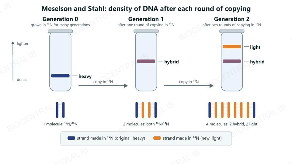
Meselson and Stahl: one band, then two

E. coli grown on heavy nitrogen (¹⁵N) were moved to light nitrogen (¹⁴N), and their DNA was spun in a density gradient after each round of copying.
  • Generation 0: one heavy band (¹⁵N/¹⁵N). Generation 1: one band of intermediate, hybrid density (¹⁵N/¹⁴N).
  • Generation 2: two bands, hybrid and light (¹⁴N/¹⁴N), in equal amounts.
  • Generation 1 rules out conservative replication (it predicts a heavy and a light band); generation 2 rules out dispersive replication (it predicts one band again, only lighter).
Why it mattersEach result was predicted in advance by one model only — this is how the experiment chose between three hypotheses.
Three centrifuge tubes. Generation 0, grown in heavy nitrogen-15: one heavy band. Generation 1, after one round of copying in nitrogen-14: one hybrid band. Generation 2, after two rounds: a hybrid band and a light band. Below each tube the DNA molecules are drawn as pairs of strands: one heavy molecule, then two hybrid molecules, then two hybrid and two light molecules
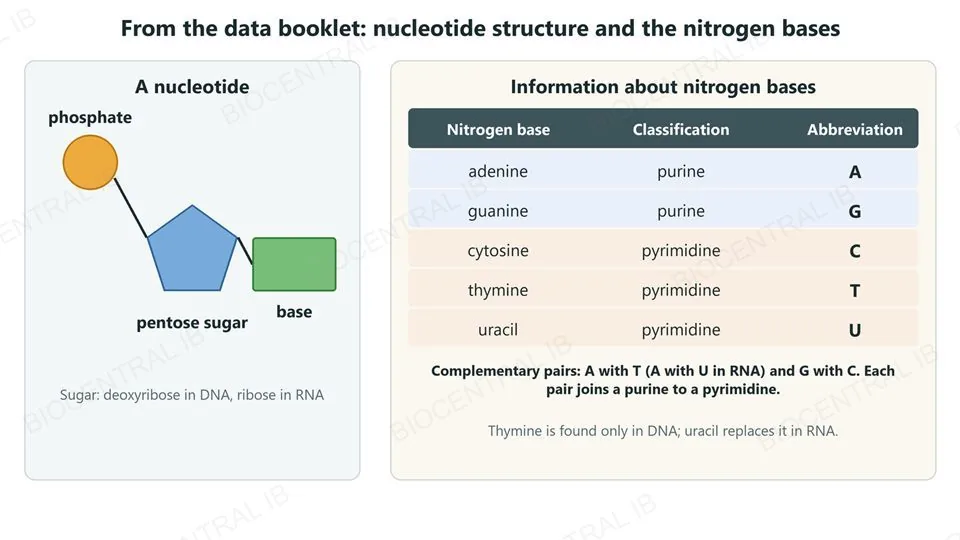
From the data booklet: a nucleotide drawn as a phosphate joined to a pentose sugar joined to a base, and a table of the nitrogen bases — adenine and guanine are purines; cytosine, thymine and uracil are pyrimidines, abbreviated A, G, C, T and U. Complementary pairs, A with T (A with U in RNA) and G with C, each join a purine to a pyrimidine
D1.1.2 · Data booklet

The bases and their pairs are in the data booklet

The data booklet shows a nucleotide as phosphate + pentose sugar + base, and lists the five nitrogen bases as purines or pyrimidines.
  • Purines: adenine (A) and guanine (G). Pyrimidines: cytosine (C), thymine (T) and uracil (U).
  • Complementary pairs, A with T and G with C, each join a purine to a pyrimidine, so every base pair is the same width.
  • Thymine is only in DNA; in RNA, uracil takes its place and pairs with A.
Why it mattersYou can check the bases in the exam — what you need is to use the pairing rules, for example to work out a complementary strand.
D1.1.3

Unwinding before copying begins

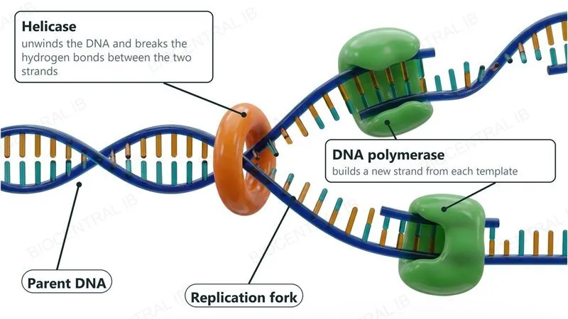
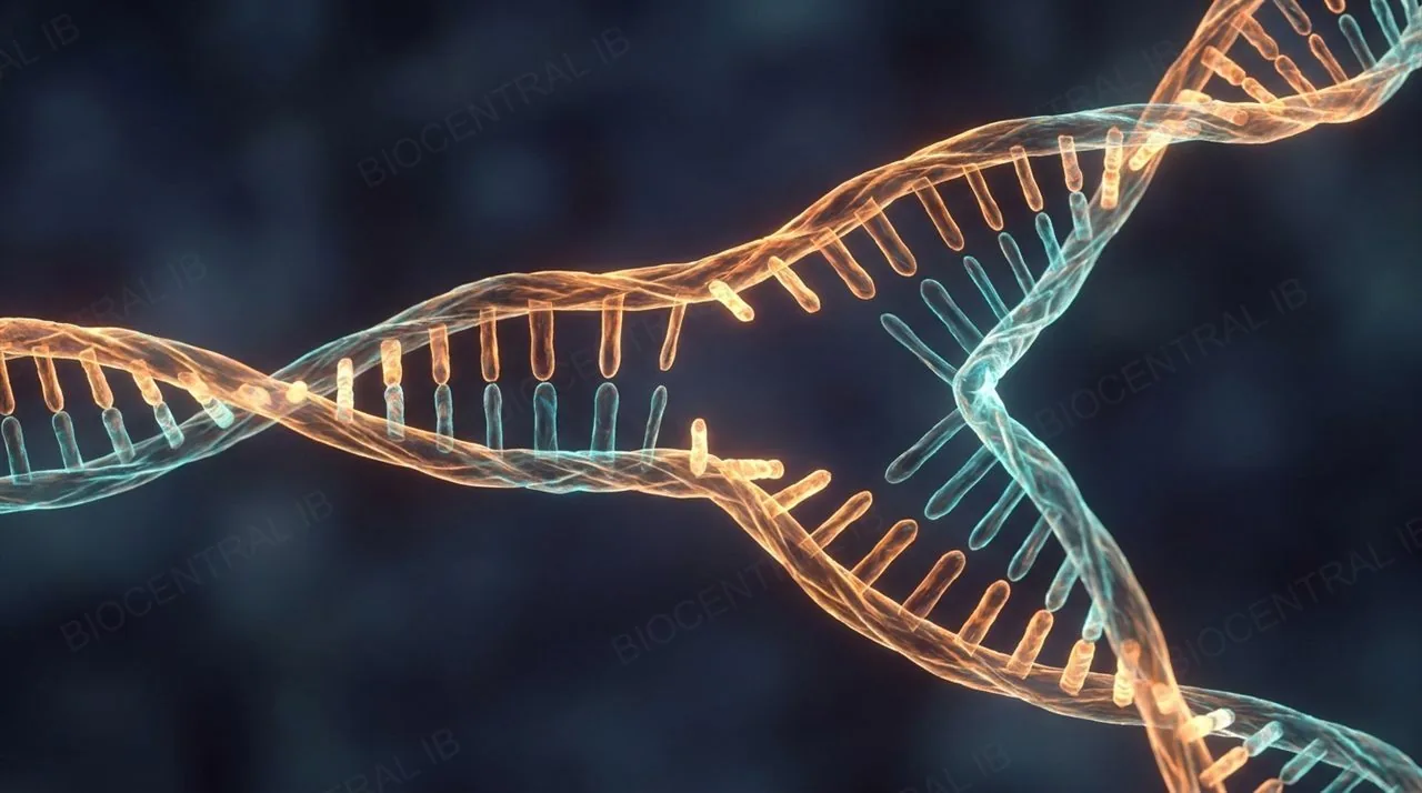
Before copying starts, the antiparallel double helix from A1.2 has to be opened: helicase unwinds the DNA, exposing each strand as a template.
  • Helicase moves along the molecule breaking the hydrogen bonds between complementary bases, separating the two strands and creating a Y-shaped replication fork.
  • DNA polymerase is the general name for the enzyme that then builds a new strand, adding nucleotides one at a time to match the exposed template.
  • At this level, it is enough to know DNA polymerase's general function — synthesizing new DNA from a template — without yet distinguishing its specific forms.
Why it mattersThis general picture is what SL students need; the specific enzymes and the leading/lagging-strand complications are additional HL content, covered later in this lesson.
A DNA double helix opening into a replication fork: helicase, an orange ring at the junction, unwinds the DNA and breaks the hydrogen bonds, and two green DNA polymerase enzymes each build a new strand on a template
Part two

PCR and gel electrophoresis — DNA in the lab

D1.1.4 – D1.1.5
D1.1.4

Copying DNA outside a cell

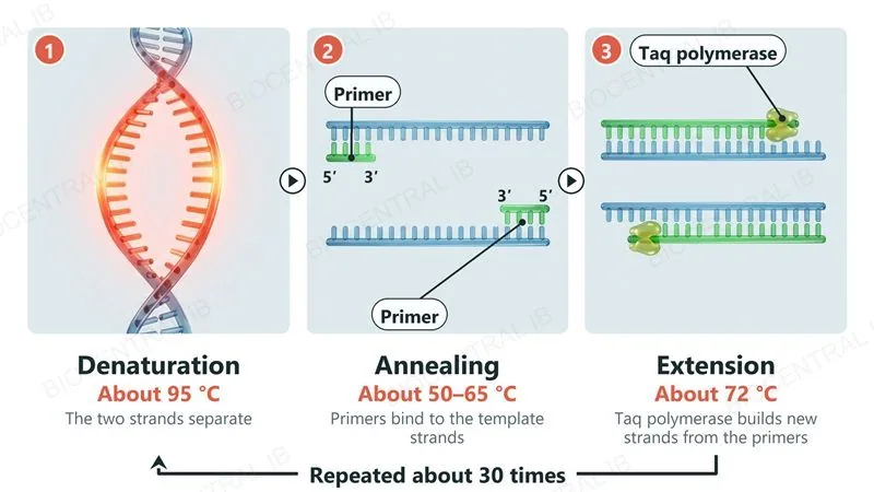
The polymerase chain reaction (PCR) is a laboratory technique that amplifies a chosen section of DNA, producing millions of copies from one starting sample.
  • A reaction mix of the DNA sample, primers (short sequences marking the target region's start and end) and Taq polymerase is cycled through set temperatures.
  • Heating separates the DNA strands, cooling lets the primers bind, and warming lets Taq polymerase extend a new strand from each primer.
  • Each cycle doubles the copy number, so repeating this three-step cycle around 30 times turns a single molecule into over a billion copies.
Why it mattersTaq polymerase, first isolated from a hot-spring bacterium, survives repeated heating that would destroy an ordinary enzyme — exactly why PCR works.
Three steps of PCR shown as photographic renders: 1 denaturation at about 95 degrees Celsius separates the strands, 2 annealing at about 50 to 65 degrees lets primers bind, 3 extension at about 72 degrees lets Taq polymerase build new strands from the primers; the cycle is repeated about 30 times
D1.1.4

Sorting DNA by size

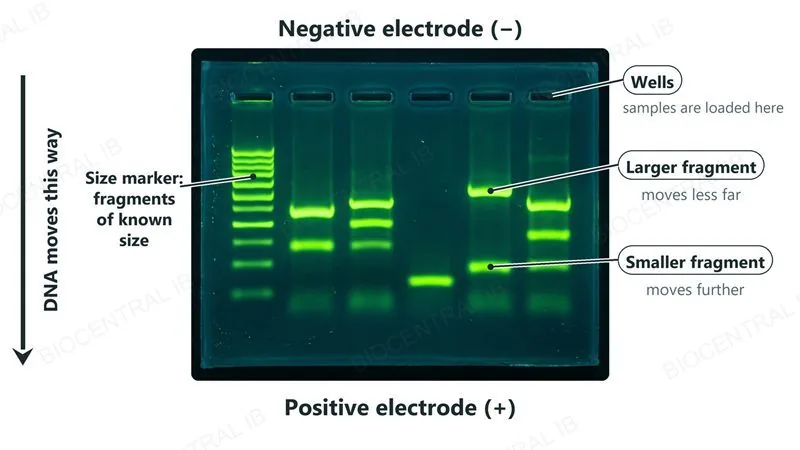
Gel electrophoresis separates DNA fragments by size: DNA's phosphate backbone is negatively charged, so an electric current pulls every fragment through the gel toward the positive electrode.
  • Samples are loaded into wells at the negative end of a porous agarose gel; when the current is switched on, the fragments travel through the gel toward the positive electrode.
  • Smaller fragments slip through the gel's pores more easily and travel further, while larger fragments are held back, so fragments separate into bands ordered by size.
  • Comparing the pattern against a size marker — a lane of fragments of known sizes — lets an unknown fragment's size be read directly off the gel.
Why it mattersPCR and gel electrophoresis are usually paired: PCR makes enough copies to see, and the gel shows whether a fragment of the expected size is present.
A photograph of an agarose gel under ultraviolet light with a lane of size-marker fragments and sample lanes; the negative electrode is at the top with the wells, the positive electrode at the bottom, and DNA moves down toward it, with larger fragments moving less far than smaller ones
D1.1.5

One technique, many uses

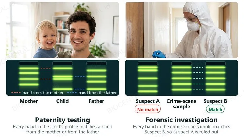
PCR and gel electrophoresis together support real applications from forensic science to paternity testing.
  • DNA profiling compares band patterns from PCR-amplified regions that vary between individuals, matching a suspect's DNA to a crime-scene sample or confirming a biological relationship.
  • Testing more separate markers sharply lowers the probability that two unrelated people would match by chance at every one of them, which is why real forensic profiles use many markers, not just one or two.
  • The same PCR-based approach also underlies rapid infectious-disease tests and prenatal genetic testing.
Why it mattersThis is a real nature-of-science example: a test's reliability improves as more independent measurements — here, markers — are combined.
Two DNA profiling uses, each with a photograph and a gel band pattern: paternity testing, where every band of a child matches a band from the mother or the father, and a forensic investigation, where the crime-scene sample matches Suspect B but not Suspect A
Quick check

A forensic lab matches a suspect's DNA to a crime-scene sample using only one genetic marker. A colleague suggests testing several more markers before reporting a positive identification. Why is this good practice?

Testing more markers reduces the probability that the match happened by chance between two unrelated people
More markers make Taq polymerase work at a lower temperature
A single marker can't be amplified by PCR at all
Gel electrophoresis can only separate two fragments at once
Correct answer: more markers reduce the chance of a false match. Increasing the number of independent markers tested sharply lowers the probability that two unrelated people would match by chance at every one of them — the same reliability principle behind repeating measurements in any experiment.
Part three

HL — the replication fork in detail

D1.1.6 – D1.1.9
D1.1.6 · HL

DNA polymerase only builds one way

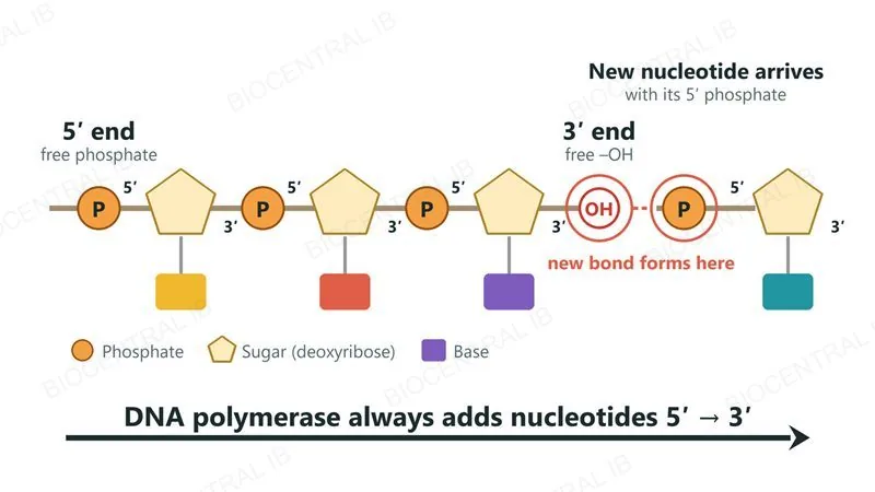
Every DNA strand has a direction, marked by its 5' end (a free phosphate group) and 3' end (a free hydroxyl group) — DNA polymerase can extend a strand in only one of these directions.
  • DNA polymerase always joins a new nucleotide's 5' phosphate onto the 3'-OH end of the growing strand, so every new strand grows from its 5' end toward its 3' end.
  • Because the two parent strands are antiparallel (running in opposite directions, as in A1.2), their 3' ends point in opposite directions at the fork.
  • This one fixed rule — always 5' to 3' — forces the two strands to be replicated by genuinely different processes, covered next.
Why it mattersThis single directional rule explains almost everything else distinctive about replication at the fork.
A DNA strand drawn as phosphate, sugar and base units with its 5 prime end, a free phosphate, on the left and its 3 prime end, a free hydroxyl group, on the right, and a new nucleotide about to join the 3 prime end through its 5 prime phosphate; DNA polymerase always adds nucleotides 5 prime to 3 prime
D1.1.7 · HL

One strand smooth, one strand in pieces

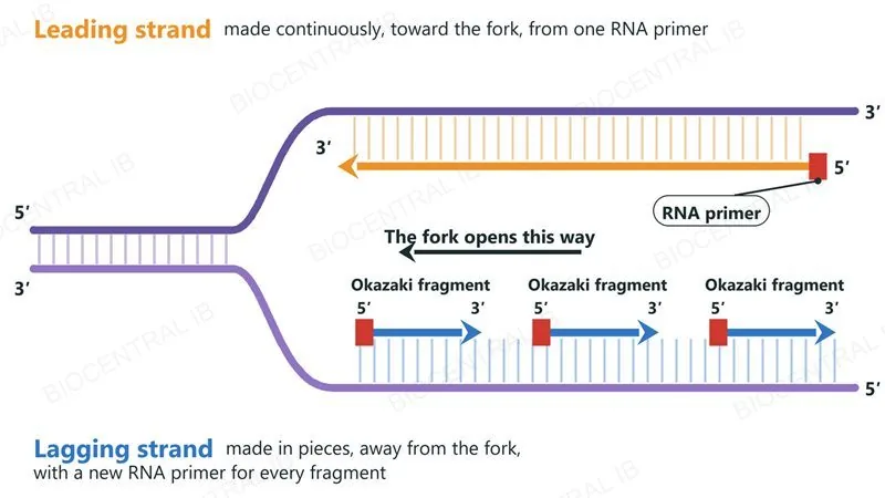
Because DNA polymerase only builds 5' to 3', and the two templates run in opposite directions, the two new strands at a fork are made by different processes.
  • The leading strand is synthesized continuously, in the same direction the fork is opening, needing only one RNA primer (a short starter sequence) to begin.
  • The lagging strand is synthesized discontinuously, in short Okazaki fragments: its template runs the other way, so DNA polymerase has to work away from the fork, copying only the stretch just exposed — a new primer is needed for each one.
  • Both strands are still built 5' to 3' overall; only the direction relative to the fork, and how often priming restarts, differs.
Why it mattersThe difference comes entirely from a fixed 5'-to-3' rule meeting antiparallel strands, not from different enzymes.
A replication fork with every 5 prime and 3 prime end labelled: the leading strand is made continuously toward the fork from one RNA primer, and the lagging strand is made away from the fork as three separate Okazaki fragments, each starting with its own RNA primer
D1.1.8 · HL

Starting and extending each strand

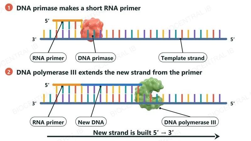
In the prokaryotic system, replication depends on a defined set of enzymes, each with a distinct job — starting with DNA primase and DNA polymerase III.
  • DNA primase synthesizes the short RNA primer needed to start each new strand, since DNA polymerase cannot begin a strand from scratch and can only extend an existing 3' end.
  • DNA polymerase III is the main replication enzyme, rapidly extending each new strand from its primer by adding nucleotides 5' to 3' along the template.
  • On the lagging strand, this primase-then-polymerase-III pairing must act repeatedly, once per Okazaki fragment, rather than just once.
Why it mattersThese functions are limited to the prokaryotic system at this level — eukaryotic replication enzymes are not required.
Step 1: DNA primase makes a short RNA primer on the template strand. Step 2: DNA polymerase III extends the new DNA strand from the primer, always 5 prime to 3 prime
D1.1.8 · HL

Removing primers, sealing the gaps

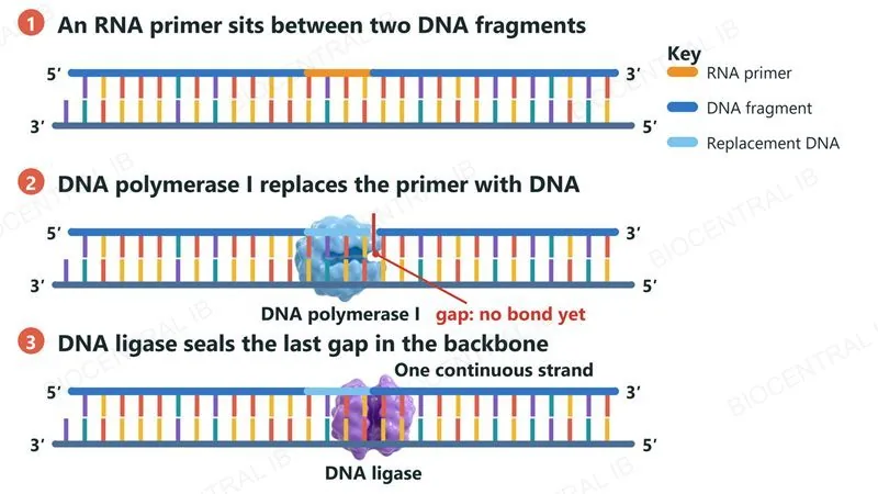
Two further prokaryotic enzymes finish what DNA primase and DNA polymerase III start: DNA polymerase I and DNA ligase.
  • DNA polymerase I removes each RNA primer and replaces it with DNA nucleotides, extending the adjacent fragment until the primer's gap is filled.
  • Because DNA polymerase I cannot join the final fragment to the strand ahead of it, DNA ligase catalyzes the last covalent bond, sealing the sugar-phosphate backbone into one continuous strand.
  • On the lagging strand this removal-and-sealing step happens once at every Okazaki-fragment boundary, joining many separate pieces into a single molecule.
Why it mattersWithout DNA ligase, the lagging strand would stay a series of disconnected fragments rather than one complete new strand.
Three steps: an RNA primer lies between two DNA fragments; DNA polymerase I replaces the primer with DNA, leaving one gap in the backbone; DNA ligase seals the gap so the new strand is one continuous piece
D1.1.7–8 · HL

Five enzymes work together at the replication fork

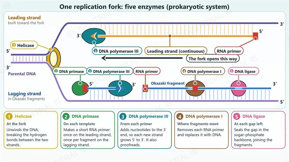
In the prokaryotic system, five enzymes copy the DNA at each fork, each with one job.
  • Helicase unwinds the DNA at the fork, breaking the hydrogen bonds between the strands.
  • DNA primase makes an RNA primer, and DNA polymerase III extends it 5′ to 3′: continuously on the leading strand, in Okazaki fragments on the lagging strand.
  • DNA polymerase I replaces each RNA primer with DNA, then DNA ligase seals the gap between fragments.
Why it mattersThe order to recall is unwind, prime, extend, replace, seal.
A labelled diagram of one prokaryotic replication fork. The parental DNA duplex on the left is unwound by a numbered helicase ring at the fork, which opens towards the left. The leading strand, on top, is built continuously toward the fork from a single RNA primer at its 5 prime end, with DNA polymerase III at its growing 3 prime end. The lagging strand, below, is built away from the fork in Okazaki fragments: DNA primase lays a new RNA primer, DNA polymerase III extends the newest fragment, DNA polymerase I replaces an older RNA primer with DNA, and DNA ligase seals the last gap so the fragments become one strand. Five numbered cards below give where and how each enzyme works: 1 helicase unwinds the DNA and breaks hydrogen bonds, 2 primase makes an RNA primer, 3 DNA polymerase III adds nucleotides to the 3 prime end and proofreads, 4 DNA polymerase I replaces the primer with DNA, 5 DNA ligase seals the gap in the backbone.
D1.1.9 · HL

Catching mistakes as they happen

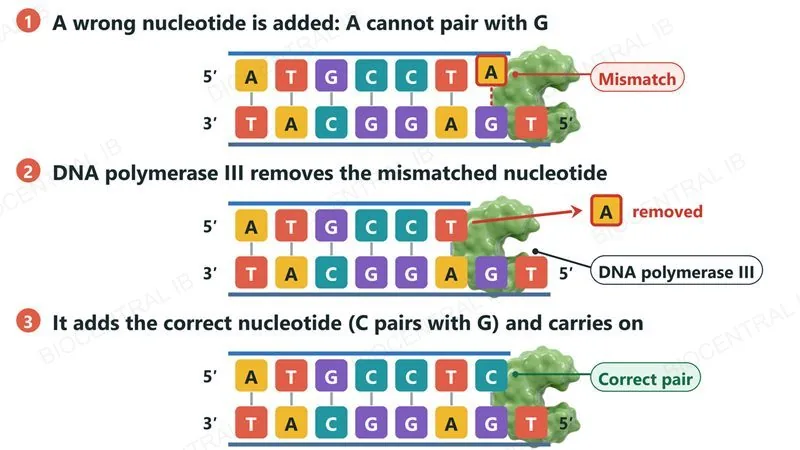
DNA polymerase III also proofreads its own work, checking each newly added nucleotide against its template as replication proceeds.
  • If a nucleotide with a mismatched base is added at the 3' terminal of the growing strand, DNA polymerase III recognizes the incorrect pairing and removes that nucleotide.
  • It then adds the correctly matched nucleotide in its place before continuing synthesis, so most errors are corrected within moments of being made.
  • This proofreading step, alongside the specificity of complementary base pairing itself, is a major reason replication achieves such high accuracy.
Why it mattersErrors that escape proofreading can become permanent mutations, passed on to every future copy of that DNA molecule.
Three steps: DNA polymerase III adds a wrong nucleotide, A opposite G, which is a mismatch; it removes that nucleotide; it then adds the correct nucleotide, C, which pairs with G, and carries on
Quick check · HL

At a single replication fork, one new strand is made in one long continuous piece while the other is made as several short fragments joined together afterwards. What causes this difference?

DNA polymerase only synthesizes 5' to 3', and the two antiparallel templates force one new strand to be built away from the fork in short bursts
The two strands are copied by completely different, unrelated enzymes with different accuracy
One strand is made of DNA and the other is temporarily made of RNA
The lagging strand's template is chemically different from the leading strand's template
Correct answer: a fixed 5'-to-3' rule meeting antiparallel strands. Because DNA polymerase can only extend a strand 5' to 3', and the two templates run in opposite directions, the leading strand is built continuously toward the fork while the lagging strand is built discontinuously, away from the fork, as Okazaki fragments.

Key vocabulary

Worth being able to define in a single sentence each

Semi-conservative replication
A model of DNA replication in which each new molecule keeps one original strand and one newly synthesized strand.
Primer
A short nucleotide sequence that provides the free 3' end DNA polymerase needs before it can begin extending a strand.
Taq polymerase
A heat-stable DNA polymerase, originally from a hot-spring bacterium, used to extend new strands during PCR.
DNA profiling
Comparing PCR-amplified, individually variable DNA regions to identify or match a person, e.g. in forensics or paternity testing.
Okazaki fragmentHL
One of the short DNA sections making up the discontinuously synthesized lagging strand.
DNA proofreadingHL
DNA polymerase III's removal and correction of a mismatched nucleotide immediately after it is added.

Where this shows up again

B1.1
DNA replication requires a supply of nucleotides. Explain how the deoxyribose sugar (B1.1) differs from ribose and why this is essential for DNA stability.
A1.1
The two strands of DNA are held together by hydrogen bonds between complementary bases. How do the properties of hydrogen bonds (A1.1) enable both stability and the separation required for replication?
D2.1
DNA replication occurs before cell division (D2.1). Explain why replication must be completed before mitosis begins, and what would happen if a cell divided with partially replicated DNA.
D1.3
Mutations (D1.3) can arise from errors in DNA replication that escape proofreading. Explain how a single base substitution during replication can lead to a change in the polypeptide sequence.

D1.1 DNA replication — one-page recap

Screenshot this slide to revise from

Why replicate
  • DNA replication produces two exact copies — needed for growth, tissue replacement and reproduction.
  • It copies rather than mixes alleles; rare errors that are not corrected become mutations (D1.3).
Semi-conservative model
  • Each new molecule = one original strand + one new strand; complementary base pairing gives high accuracy.
  • Meselson–Stahl's density-gradient experiment provided the classic evidence for this model.
Helicase & DNA polymerase
  • Helicase unwinds the double helix, breaking hydrogen bonds between strands.
  • DNA polymerase builds a new strand from a template (SL: general function only).
PCR & gel electrophoresis
  • PCR amplifies DNA using primers, Taq polymerase and repeated heating/cooling cycles.
  • Gel electrophoresis separates fragments by size using an electric current; more markers tested = lower chance of a false match.
HL · Leading & lagging strands
  • DNA polymerase only builds 5'→3': the leading strand is continuous, the lagging strand is discontinuous (Okazaki fragments).
  • Primase starts each fragment; polymerase III extends it; polymerase I replaces primers; ligase seals the gaps.
HL · Proofreading
  • DNA polymerase III proofreads and replaces mismatched nucleotides as it goes, boosting accuracy further.
  • Errors that escape proofreading can become permanent mutations (D1.3).
Next step

Now test yourself on D1.1

Reading the slides is half the job. The other half is answering questions the way the exam asks them.

39 quiz questions6 data questions5 exam questionsmarkschemes included

Unlock every lesson with Pro — quizzes, data questions and exam papers. Two lessons are completely free to try: A1.1 Water and B1.1 Carbohydrates and lipids.

One-page summary, key terms and sample questions for D1.1 →

Copied, checked, and ready to be read

From one double helix to two identical copies — the same enzymes that keep every cell's genome intact now power the tools that can identify a person from a single sample.
D1.1 DNA replication · BioCentral IB
Use ↓ ↑ or click to navigate
01 / 24