BioCentral IBBioCentral IB
IB Biology · Theme B · B3.3 · HL only

Muscle and motility

How muscles contract, how skeletons and joints turn contraction into movement, and why animals move. The whole topic is HL only.
Guiding questions

How do muscles contract and cause movement?

What are the benefits to animals of having muscle tissue?

Part one · HL

Movement and muscle contraction

B3.3.1 – B3.3.4
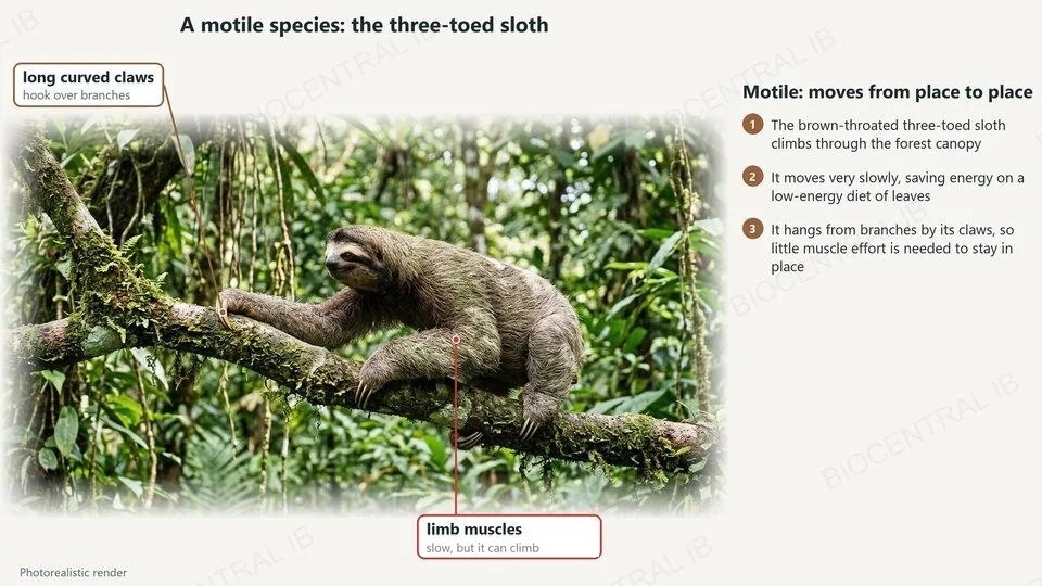
A photorealistic render of a brown-throated three-toed sloth climbing along a branch, labelled: long curved claws and limb muscles
B3.3.1 · HL

A motile species: the sloth

Movement is a universal feature of living organisms. A motile species can move from place to place.
  • The brown-throated three-toed sloth climbs through the rainforest canopy.
  • It moves very slowly, which saves energy: its diet of leaves provides little.
  • Long curved claws hook over branches, so it can hang with little muscle effort.
RememberSlow is still motile: the sloth moves from place to place.
B3.3.1 · HL

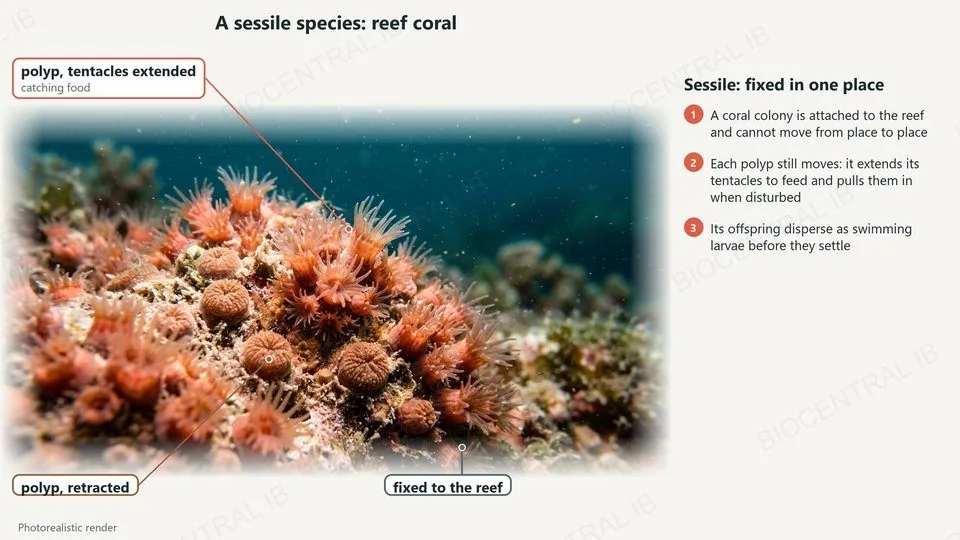
A sessile species: coral

A sessile species is fixed in one place, but it still moves.
  • A reef-building coral colony is attached to the reef and cannot move from place to place.
  • Each polyp extends its tentacles to catch food and pulls them in when disturbed.
  • Its offspring disperse as swimming larvae before they settle and attach.
CompareMotile: moves from place to place. Sessile: fixed, but parts move.
A photorealistic render of reef coral, labelled: a polyp with tentacles extended, a retracted polyp, and the colony fixed to the reef
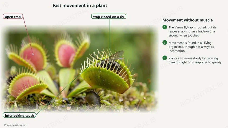
A photorealistic render of Venus flytraps, labelled: an open trap, a trap closed on a fly, and the interlocking teeth
B3.3.1 · HL

Movement in a plant

Plants are rooted, but they move too.
  • A Venus flytrap’s leaves snap shut in a fraction of a second when an insect touches the hairs inside.
  • Most plant movement is slower: growing towards light or in response to gravity.
  • Movement happens in all living organisms, even when it is not locomotion.
RememberNot every organism moves from place to place, but all show movement.
B3.3.2 · HL

Inside a muscle

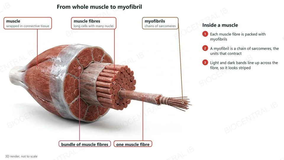
Skeletal muscle is built from long cells called muscle fibres.
  • A muscle is made of bundles of muscle fibres, wrapped in connective tissue.
  • Each fibre is a long cell with many nuclei, packed with myofibrils.
  • A myofibril is a chain of sarcomeres, the units that contract.
  • The bands of the sarcomeres line up across the fibre, so it looks striped.
LinkMuscle fibres form by fusion of cells (B2.3).
A 3D render of a skeletal muscle cut open, labelled: the muscle, a bundle of muscle fibres, muscle fibres, one muscle fibre and striped myofibrils
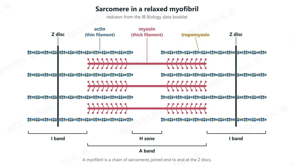
The data booklet diagram of a sarcomere in a relaxed myofibril, redrawn: Z discs, actin thin filaments with tropomyosin, myosin thick filaments, I bands, A band and H zone
B3.3.2 · Data booklet · HL

The sarcomere

A sarcomere runs from one Z disc to the next.
  • Actin (thin filaments) is attached to the Z discs, with tropomyosin running along it.
  • Myosin (thick filaments) lies in the centre, overlapping the actin.
  • A band: the length of the myosin filaments. I band: actin only. H zone: myosin only.
Data bookletThis diagram is in the data booklet; it is redrawn here.
B3.3.2 · HL

The sliding filament model

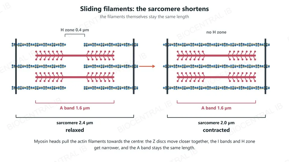
A sarcomere contracts by the sliding of actin and myosin filaments.
  • Myosin heads pull the actin filaments towards the centre of the sarcomere.
  • The Z discs move closer, so the sarcomere shortens.
  • The I band and H zone narrow; the A band stays the same length.
  • The filaments themselves do not change length: they overlap more.
CalculateShortening from 2.4 to 2.0 µm is (0.4 ÷ 2.4) × 100 ≈ 17%.
A relaxed sarcomere of 2.4 micrometres and a contracted one of 2.0 micrometres: the A band stays 1.6 micrometres, the H zone disappears and the Z discs move closer
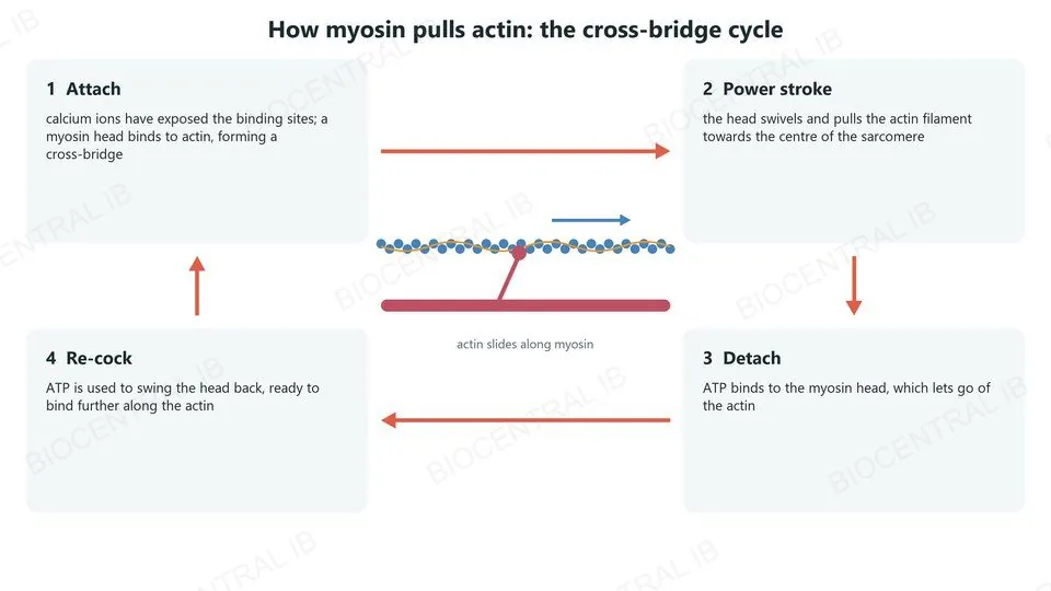
The cross-bridge cycle in four steps: attach, power stroke, detach when ATP binds, and re-cock using ATP
B3.3.2 · HL

How myosin pulls actin

Myosin heads pull actin in a repeated cycle of cross-bridges.
  • A myosin head binds to actin, forming a cross-bridge.
  • The head swivels in a power stroke, pulling the actin filament.
  • ATP binds and the head detaches; energy from ATP re-cocks it, ready to bind again further along.
RememberWithout ATP, myosin cannot detach: the muscle stays stiff.
B3.3.2 · B3.3.4 · HL

From impulse to contraction

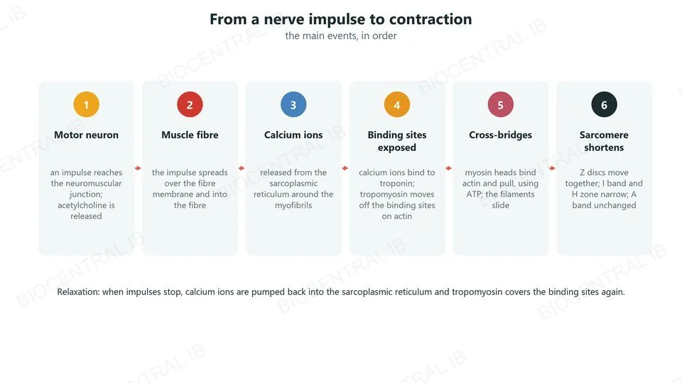
A nerve impulse becomes a shortened sarcomere through one chain of events.
  • A motor neuron releases acetylcholine at the neuromuscular junction; the impulse spreads into the fibre.
  • The sarcoplasmic reticulum releases calcium ions, which bind to troponin, so tropomyosin uncovers the binding sites on actin.
  • Myosin heads form cross-bridges and pull: the sarcomeres shorten.
  • When impulses stop, calcium ions are pumped back and the muscle relaxes.
RememberCalcium ions switch contraction on; ATP keeps it going.
Six steps from a nerve impulse to contraction: motor neuron, muscle fibre, calcium ions released, binding sites exposed, cross-bridges, sarcomere shortens
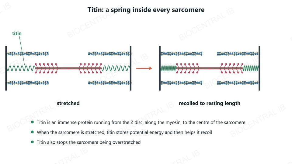
A stretched sarcomere with titin drawn as springs from the Z discs to the myosin filament, and the same sarcomere recoiled to its resting length
B3.3.3 · HL

Titin: a molecular spring

Titin is an immense protein that runs from the Z disc, along the myosin filament, to the centre of every sarcomere.
  • When a sarcomere is stretched, titin stores potential energy.
  • It then helps the sarcomere recoil to its resting length.
  • Titin also prevents the sarcomere being overstretched.
Why it mattersRecoil after stretching needs no contraction of the stretched muscle.
B3.3.3 · HL

Antagonistic muscles

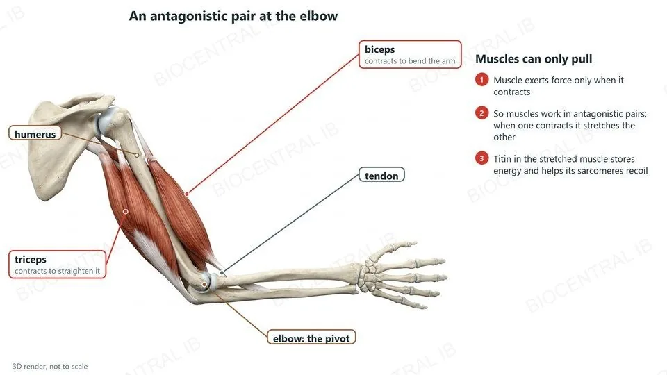
Muscle tissue can only exert force when it contracts, so muscles work in antagonistic pairs.
  • The biceps contracts to bend the arm at the elbow; the triceps contracts to straighten it.
  • When one muscle of the pair contracts, it stretches the other.
  • Titin in the stretched muscle helps its sarcomeres recoil, ready to contract again.
RememberMuscles pull; they never push.
A 3D render of the arm bones and muscles, labelled: biceps, triceps, tendon, humerus and the elbow as the pivot
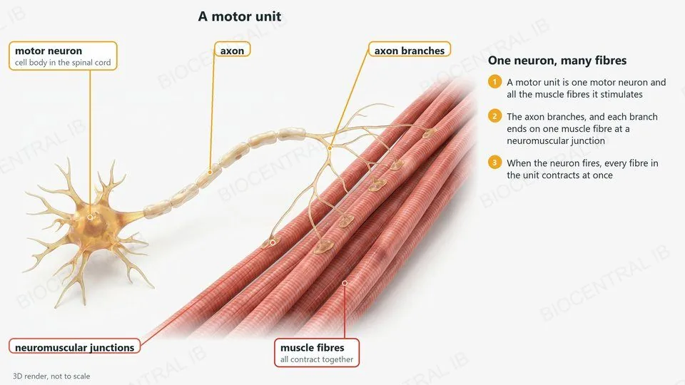
A 3D render of a motor neuron whose axon branches to several muscle fibres, labelled: motor neuron, axon, branches, neuromuscular junctions and muscle fibres
B3.3.4 · HL

A motor unit

A motor unit is one motor neuron and all the muscle fibres it stimulates.
  • The axon of the motor neuron branches near the muscle.
  • Each branch ends on one muscle fibre at a neuromuscular junction.
  • When the neuron fires, all the fibres in the unit contract together.
  • Small units (few fibres) give fine control; large units give more force.
ExampleThe muscles that move the eye have small motor units.
B3.3.4 · HL

The neuromuscular junction

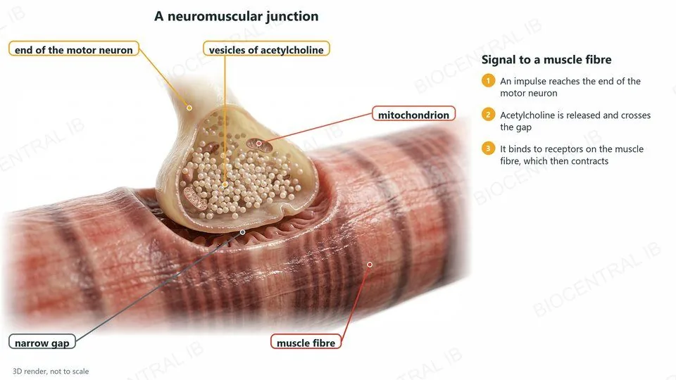
The neuromuscular junction connects the motor neuron to a muscle fibre.
  • An impulse arrives at the end of the motor neuron.
  • Vesicles release acetylcholine, which crosses the narrow gap.
  • It binds to receptors on the muscle fibre, which then contracts.
  • Each muscle fibre receives impulses from only one motor neuron.
LinkThe neuromuscular junction is a synapse (C2.2).
A 3D render of a neuromuscular junction cut open, labelled: the end of the motor neuron, vesicles of acetylcholine, a mitochondrion, the narrow gap and the muscle fibre
Quick check · HL

A sarcomere contracts. Which part stays the same length?

the I band
the A band
the H zone
the sarcomere
Correct answer: the A band. It is the length of the myosin filaments, which do not shorten. The actin slides further in, so the I band and H zone narrow and the sarcomere shortens.
Part two · HL

Skeletons, joints and the ribcage

B3.3.5 – B3.3.8
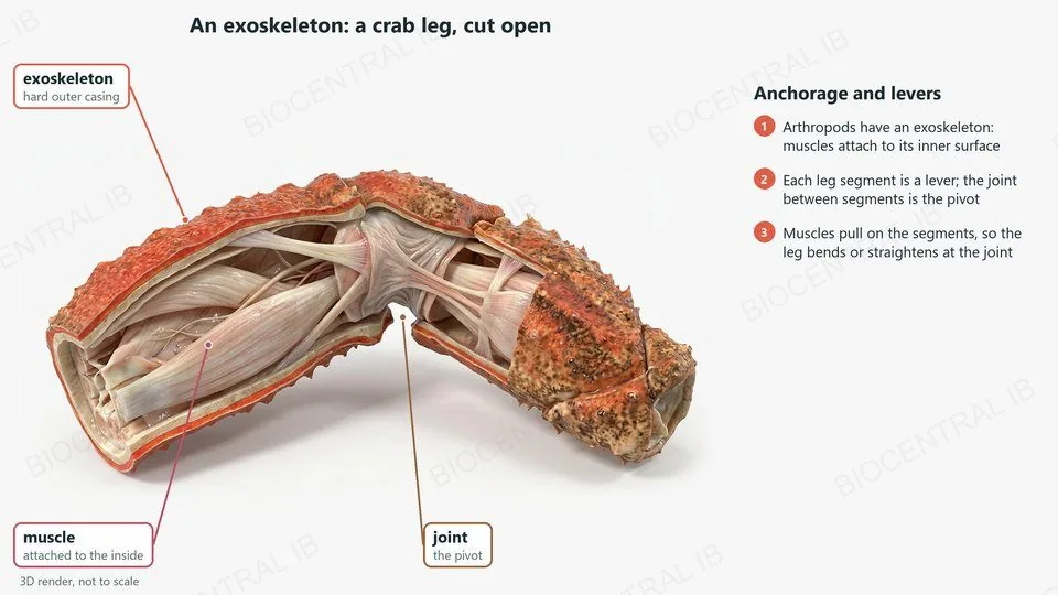
A 3D render of a crab leg cut open, labelled: the exoskeleton, muscle attached to its inside, and the joint as the pivot
B3.3.5 · HL

An exoskeleton

Arthropods, such as crabs and insects, have an exoskeleton: a hard outer casing.
  • Muscles attach to the inner surface of the exoskeleton: it is their anchorage.
  • Each segment of a leg acts as a lever, and the joint between segments is the pivot.
  • Muscles pull on the segments, so the leg bends or straightens at the joint.
CompareExoskeleton: muscles inside. Endoskeleton: muscles outside.
B3.3.5 · HL

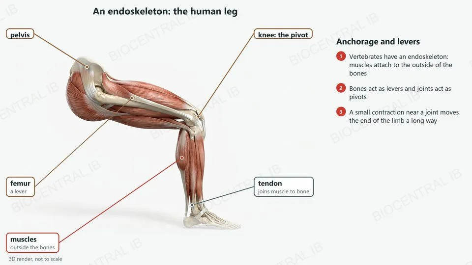
An endoskeleton

Vertebrates have an endoskeleton: bones inside the body.
  • Muscles attach to the outside of the bones by tendons.
  • Bones act as levers and joints act as pivots.
  • Muscles attach close to a joint, so a short contraction moves the end of the limb a long way.
RememberSkeletons give anchorage for muscles and act as levers.
A 3D render of a human leg, labelled: pelvis, femur as a lever, the knee as the pivot, muscles outside the bones, and a tendon
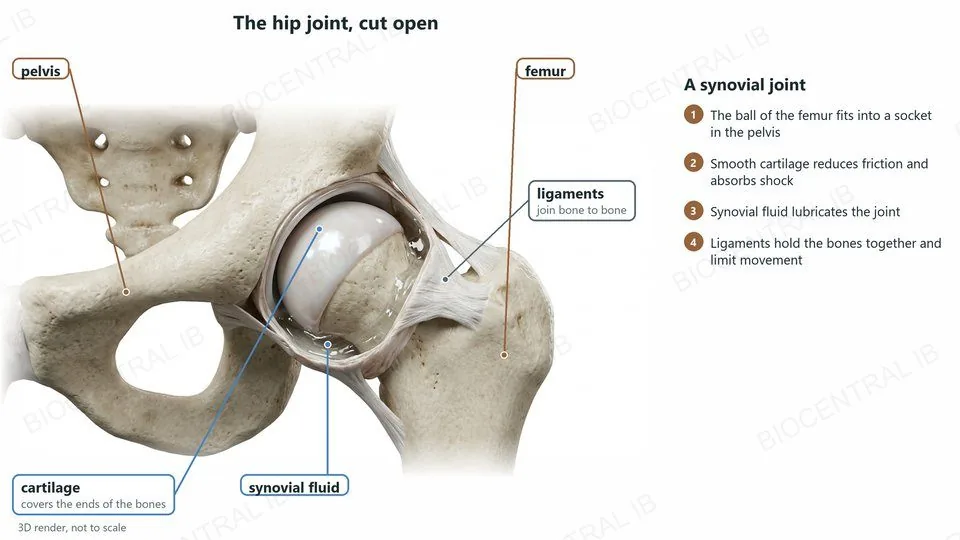
A 3D render of the hip joint cut open, labelled: pelvis, femur, cartilage, synovial fluid and ligaments
B3.3.6 · HL

The hip: a synovial joint

The hip is a synovial joint between the femur and the pelvis.
  • Bones: the ball of the femur fits into a socket in the pelvis; they act as levers.
  • Cartilage covers the ends of the bones, reducing friction and absorbing shock.
  • Synovial fluid lubricates the joint.
  • Ligaments join bone to bone, holding the joint together and limiting movement.
RememberName the femur and pelvis; named muscles and ligaments are not needed.
B3.3.6 · HL

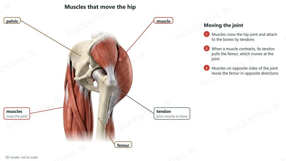
Muscles and tendons move the hip

Muscles that cross the hip joint move the femur.
  • Tendons join the muscles to the bones and transmit their pull.
  • When a muscle contracts, it pulls the femur, which moves at the joint.
  • Muscles on opposite sides of the joint move the femur in opposite directions: antagonistic pairs.
CompareLigaments: bone to bone. Tendons: muscle to bone.
A 3D render of the hip and thigh, labelled: pelvis, femur, muscles crossing the joint and a tendon
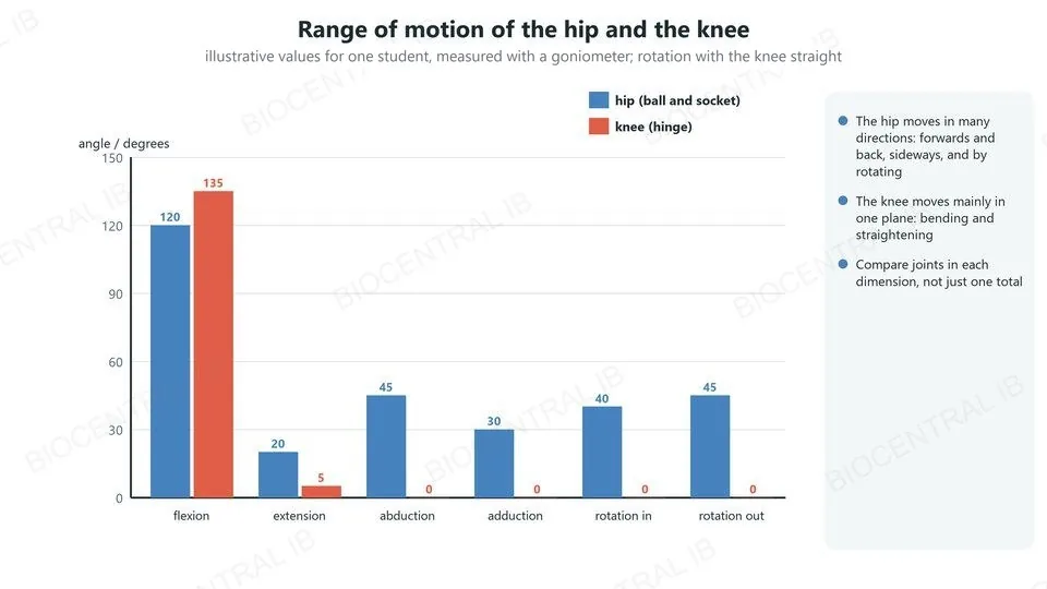
A bar chart of illustrative ranges of motion: the hip moves in six directions, the knee mainly bends and straightens
B3.3.7 · HL

Range of motion

The range of motion of a joint is how far it can move in each direction.
  • The hip, a ball-and-socket joint, moves in several dimensions: bending and straightening, sideways, and rotation.
  • The knee, a hinge joint, moves mainly in one plane: bending and straightening.
  • Compare joints in each dimension, not just as one total.
SkillCompare the range of motion of joints in a number of dimensions.
B3.3.7 · Skill · HL

Measuring a joint angle

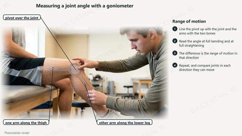
Joint angles can be measured with a goniometer or by computer analysis of images.
  • Line up the pivot of the goniometer with the joint and its arms with the two bones.
  • Read the angle at full bending and at full straightening; the difference is the range of motion.
  • Repeat the measurement and calculate a mean.
SkillImage analysis: photograph the joint and measure the angle on screen.
A photorealistic render of a student measuring the angle of a bent knee with a goniometer, labelled: the pivot over the joint and the two arms along the thigh and lower leg
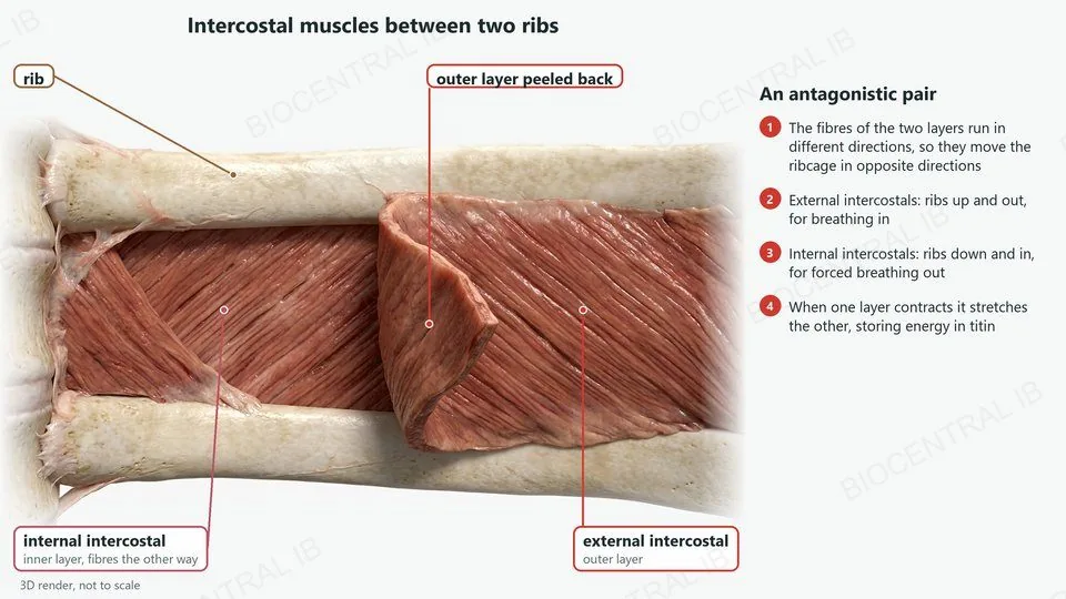
A 3D render of intercostal muscles between two ribs, labelled: rib, external intercostal, internal intercostal with fibres the other way, and the outer layer peeled back
B3.3.8 · HL

The intercostal muscles

The internal and external intercostal muscles are an antagonistic pair that moves the ribcage.
  • The fibres of the two layers run in different directions, so they move the ribcage in opposite directions.
  • External intercostals move the ribs up and out (breathing in); internal intercostals move them down and in (forced breathing out).
  • When one layer contracts, it stretches the other, storing potential energy in titin.
LinkVentilation of the lungs is in B3.1.
Quick check · HL

Which structure joins a muscle to a bone?

a ligament
a tendon
cartilage
synovial fluid
Correct answer: a tendon. Tendons transmit the pull of a muscle to a bone. Ligaments join bone to bone; cartilage and synovial fluid reduce friction in the joint.
Part three · HL

Why and how animals move

B3.3.9 – B3.3.10
B3.3.9 · HL

Moving to find food

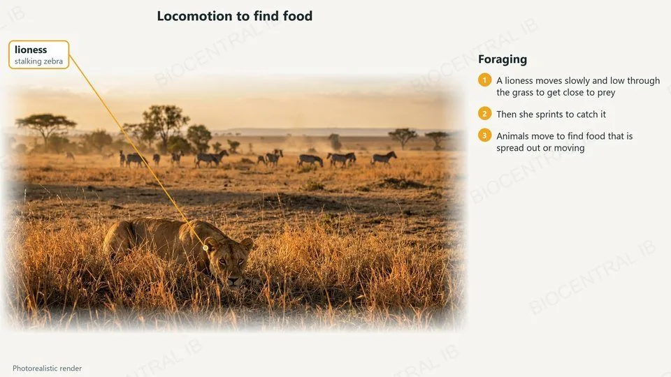
Animals move for four main reasons; the first is foraging for food.
  • A lioness stalks slowly and low through the grass to get close to zebra.
  • Then she sprints a short distance to catch one.
  • Food is often spread out or moving, so animals must travel to find it.
ExampleForaging: a lioness hunting zebra.
A photorealistic render of a lioness stalking zebra in long grass
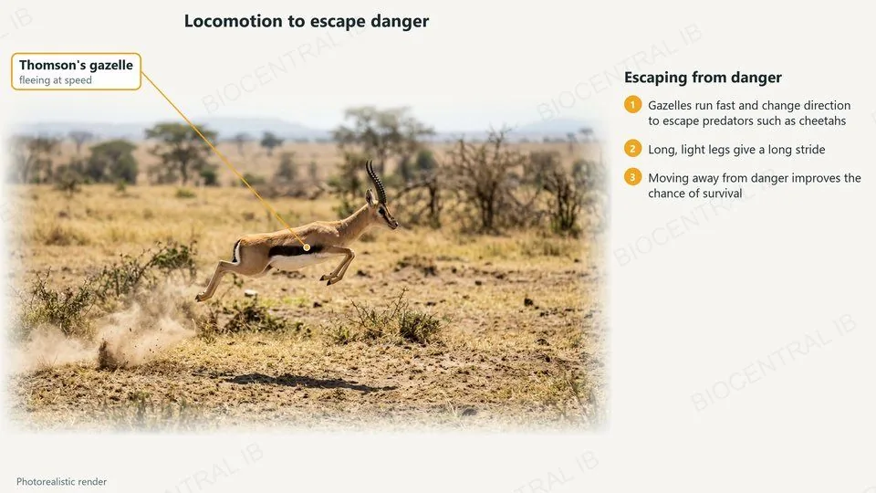
A photorealistic render of a Thomson's gazelle leaping as it flees
B3.3.9 · HL

Moving to escape danger

Locomotion lets an animal escape from danger.
  • A Thomson’s gazelle runs fast and changes direction to escape predators such as cheetahs.
  • Long, light legs give it a long stride.
  • Moving away from danger improves its chance of survival.
  • Many prey animals flee in herds, so a predator has trouble picking one out.
ExampleEscaping danger: a gazelle fleeing a predator.
B3.3.9 · HL

Moving to find a mate

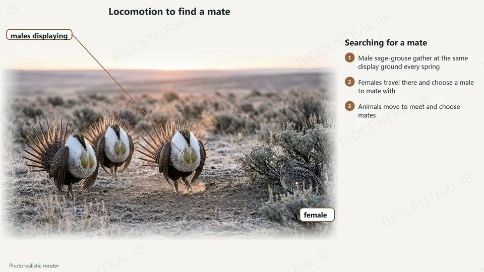
Locomotion lets animals search for a mate.
  • Male sage-grouse gather at the same display ground every spring and display.
  • Females travel to the display ground and choose a male to mate with.
  • Moving to meet allows animals to choose mates.
  • The display ground can be several kilometres from where the birds feed.
ExampleSearching for a mate: sage-grouse at a display ground.
A photorealistic render of male sage-grouse displaying at a display ground, with a female nearby
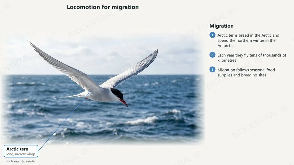
A photorealistic render of an Arctic tern flying over the sea
B3.3.9 · HL

Migration

Migration is seasonal movement between two areas.
  • Arctic terns breed in the Arctic in the northern summer.
  • They spend the northern winter in the Antarctic, where it is summer.
  • Each year they fly tens of thousands of kilometres, following food and long days.
  • Migration lets them use two places that are each good for only part of the year.
ExampleMigration: Arctic terns between the Arctic and the Antarctic.
B3.3.10 · HL

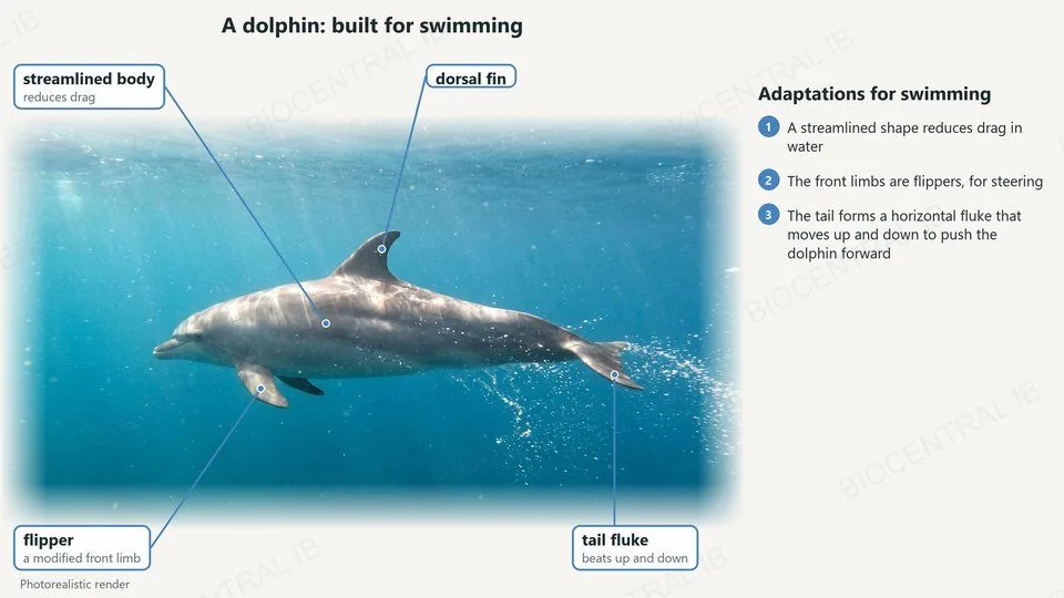
Built for swimming

Marine mammals are adapted for swimming.
  • Streamlining: a smooth, tapered body reduces drag.
  • Flippers: the front limbs are adapted to form flippers for steering.
  • Fluke: the tail forms a horizontal fluke that moves up and down to push the animal forward.
CompareFish tails move from side to side; mammal flukes move up and down.
A photorealistic render of a dolphin, labelled: streamlined body, flipper, dorsal fin and a tail fluke that beats up and down
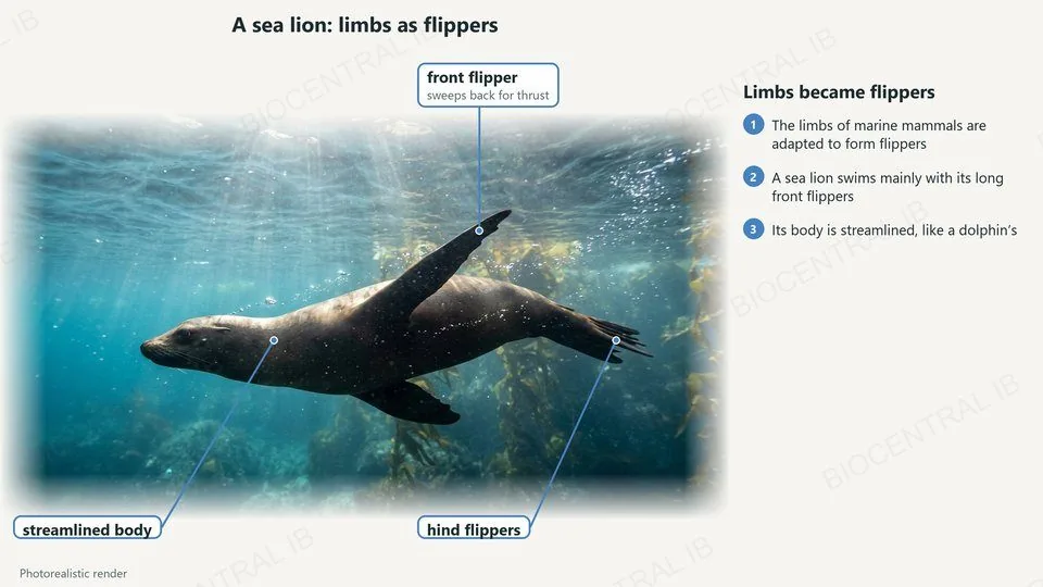
A photorealistic render of a swimming sea lion, labelled: front flipper, hind flippers and streamlined body
B3.3.10 · HL

Limbs became flippers

In marine mammals the limbs have been adapted to form flippers.
  • A sea lion swims mainly with its long front flippers, sweeping them back for thrust.
  • Its hind flippers help it steer.
  • Its body is streamlined, like a dolphin’s.
  • On land it can turn its hind flippers forward to walk.
ExampleSea lions: front flippers; dolphins: tail fluke.
B3.3.10 · HL

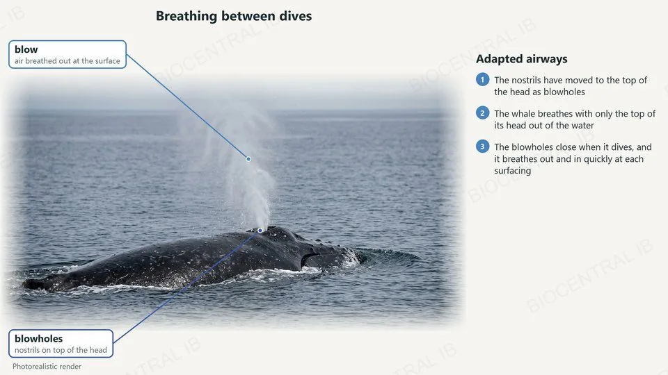
Breathing between dives

The airways of marine mammals are adapted for periodic breathing between dives.
  • In whales and dolphins the nostrils form blowholes on top of the head: one in dolphins, two in baleen whales.
  • The animal breathes with only the top of its head out of the water.
  • The blowhole closes during a dive; at the surface it breathes out and in quickly.
RememberBreathing happens only at the surface, between dives.
A photorealistic render of a humpback whale at the surface, labelled: the blow of breath and the blowholes on top of the head

Key vocabulary · HL

Worth being able to define in a single sentence each

Motile / sessile
able to move from place to place / fixed in one place.
Sarcomere
the unit of a myofibril, from one Z disc to the next.
Sliding filament model
actin slides past myosin, so the sarcomere shortens.
Cross-bridge
a myosin head bound to actin.
Titin
an elastic protein that helps sarcomeres recoil.
Antagonistic muscles
a pair in which each stretches the other.
Motor unit
one motor neuron and the fibres it stimulates.
Synovial joint
a joint with cartilage and synovial fluid, e.g. the hip.
Range of motion
how far a joint can move in each direction.

Where this shows up again

D4.1 · Natural selection
What are the advantages and disadvantages of dispersal of offspring from their parents?
D4.1 · Natural selection
In what ways does locomotion contribute to evolution within living organisms?
B3.1 · Gas exchange
How does the antagonistic action of the intercostal muscles change the volume of the thorax?
C2.2 · Neural signalling
How is the neuromuscular junction similar to a synapse between two neurons?

B3.3 Muscle and motility — one-page recap · HL

Screenshot this slide to revise from

Movement
  • All organisms move; motile (sloth) vs sessile (coral).
  • Plants move too: the Venus flytrap.
Sarcomere
  • Z disc to Z disc; actin thin, myosin thick.
  • Contraction: I band and H zone narrow, A band constant.
Contraction
  • Calcium ions expose sites; myosin heads pull actin.
  • ATP detaches and re-cocks the heads.
Relaxation and motor units
  • Titin recoils; antagonistic pairs stretch each other.
  • Motor unit: one neuron, its fibres, neuromuscular junctions.
Skeletons and joints
  • Exo vs endoskeleton: anchorage and levers.
  • Hip: cartilage, synovial fluid, ligaments, tendons; range of motion.
Locomotion
  • Food, danger, mate, migration: one example each.
  • Marine mammals: streamlined, flippers, fluke up and down, blowholes.
Next step

Now test yourself on B3.3

Reading the slides is half the job. The other half is answering questions the way the exam asks them.

47 quiz questions6 data questions5 exam questionsmarkschemes included

Unlock every lesson with Pro — quizzes, data questions and exam papers. Two lessons are completely free to try: A1.1 Water and B1.1 Carbohydrates and lipids.

One-page summary, key terms and sample questions for B3.3 →

One pull, many movements.

Sarcomeres only shorten; skeletons, joints and antagonistic pairs turn that into a sprint, a dive or a breath.
B3.3 Muscle and motility · BioCentral IB
Use ↓ ↑ or click to navigate
01 / 20