
BioCentral IBBioCentral IB
IB Biology · Theme B · B3.2 · SL and HL

Transport

How blood moves through vessels and the heart, and how water and sugars move through plants. Part one is for SL and HL; part two is HL only.
Guiding questions

What adaptations facilitate transport of fluids in animals and plants?

What are the differences and similarities between transport in animals and plants?

Part one

Blood vessels, and water in plants

B3.2.1 – B3.2.10
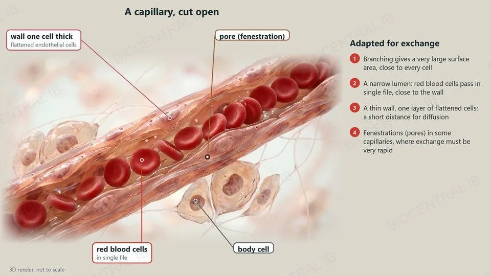
A 3D render of a capillary cut open, labelled: a wall one cell thick of flattened endothelial cells, a pore (fenestration), red blood cells in single file and a body cell
B3.2.1

Capillaries: built for exchange

Capillaries exchange materials between the blood and the internal or external environment.
  • Branching: capillaries form dense networks with a large surface area, so every cell is close to one.
  • Narrow diameter: red blood cells pass almost in single file, close to the wall.
  • Thin walls: one layer of flattened endothelial cells gives a short diffusion distance.
  • Fenestrations: some capillaries have pores in their walls, where exchange must be particularly rapid.
ExampleFenestrated capillaries in the kidney let fluid leave the blood quickly.
B3.2.2 · Skill

Arteries and veins in a micrograph

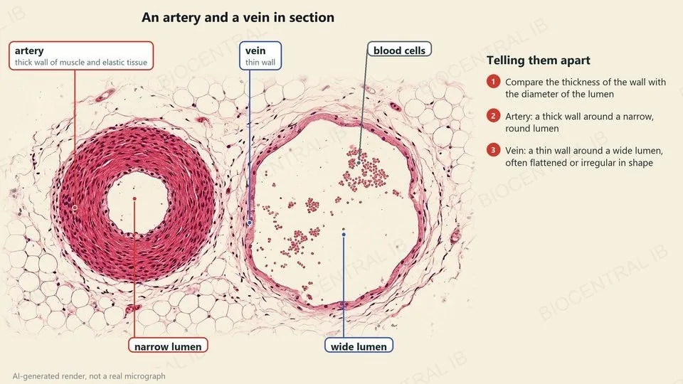
Arteries and veins can be told apart in a micrograph by the thickness of the wall relative to the diameter of the lumen.
  • Artery: a thick wall of muscle and elastic tissue around a narrow, round lumen.
  • Vein: a thin wall around a wide lumen; it is often flattened or irregular in shape.
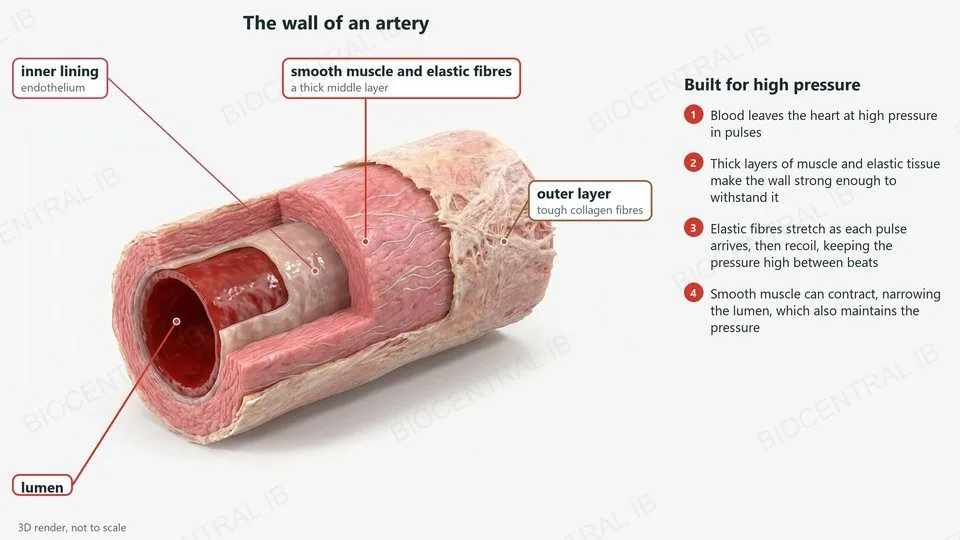
  • Both walls have the same layers: an inner lining of endothelium, a middle layer of muscle and elastic fibres, and a tough outer layer.
SkillJudge wall thickness against the lumen, not the size of the whole vessel.
An AI-generated render, not a real micrograph, of an artery and a vein in section: the artery has a thick wall of muscle and elastic tissue around a narrow lumen; the vein has a thin wall around a wide lumen
A 3D cutaway render of an artery wall, labelled: inner lining (endothelium), a thick middle layer of smooth muscle and elastic fibres, a tough outer layer of collagen, and the lumen
B3.2.3

Arteries: walls for high pressure

Arteries carry blood away from the heart at high pressure, in a surge with each heartbeat.
  • Thick layers of muscle and elastic tissue make the wall strong enough to withstand the high pressure.
  • Elastic fibres stretch as each surge of blood arrives, then recoil, so the pressure stays high between beats and the flow is steadier.
  • Smooth muscle can contract, narrowing the lumen, which also helps to maintain the pressure.
Why it mattersElastic recoil is why arterial pressure does not fall to zero between beats.
B3.2.4 · Skill

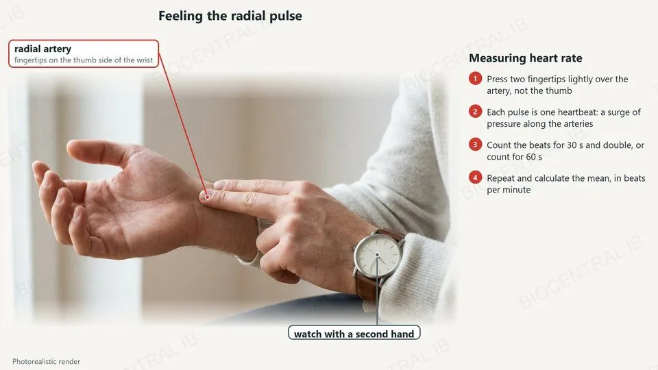
Feeling the radial pulse

Heart rate can be measured by feeling a pulse with the fingertips.
  • Each pulse is one heartbeat: a surge of pressure travelling along the arteries.
  • The radial pulse is felt on the thumb side of the wrist, where the artery runs over bone.
  • Press lightly with two fingertips, not the thumb, which has its own pulse.
  • Count the beats for 30 s and double, or for 60 s; repeat and calculate the mean in beats per minute.
Calculate18 beats in 15 s × 4 = 72 beats per minute.
A photorealistic render of two fingertips feeling the radial pulse on the thumb side of the wrist, with a watch with a second hand
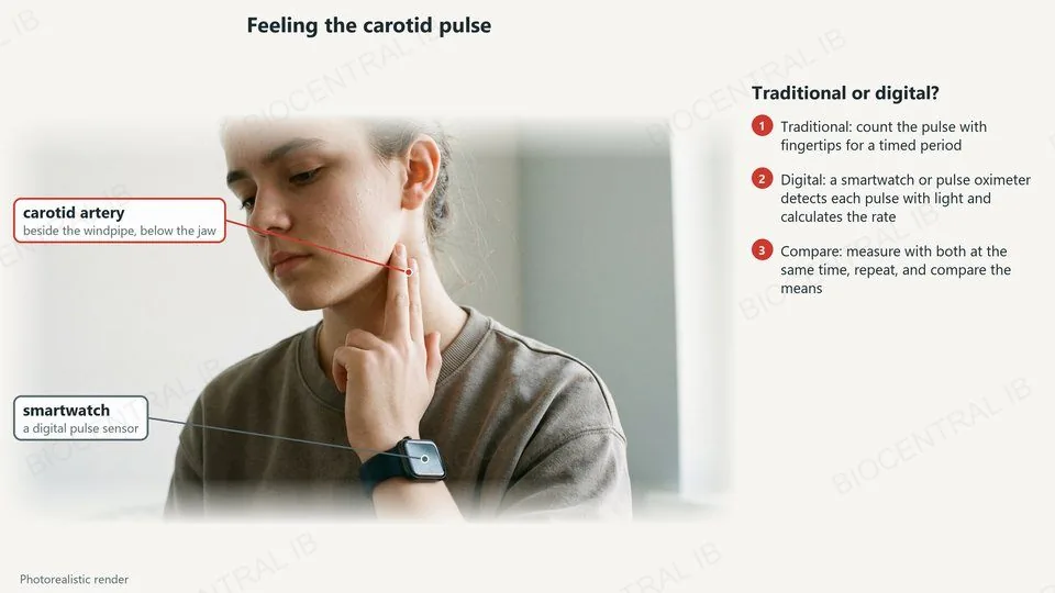
A photorealistic render of a teenager feeling the carotid pulse beside the windpipe, wearing a smartwatch with a digital pulse sensor
B3.2.4 · Skill

The carotid pulse, traditional or digital

The carotid pulse is felt in the neck, beside the windpipe and just below the jaw.
  • Traditional: count the pulse with the fingertips over a timed period.
  • Digital: a smartwatch or pulse oximeter detects each pulse with light and calculates the rate.
  • Compare: measure with both methods at the same time, repeat, and compare the means.
  • Digital methods avoid counting errors and can record continuously; their accuracy still depends on the sensor fitting well.
RememberPress gently on one side of the neck only.
B3.2.5

Veins: returning blood to the heart

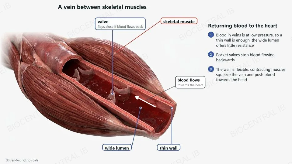
Veins return blood to the heart at low pressure.
  • A thin wall is enough at low pressure, and the wide lumen offers little resistance to flow.
  • Valves: pocket valves close if blood starts to flow backwards, preventing backflow.
  • Flexible walls: when nearby skeletal muscles contract they compress the vein, and the valves make sure the blood is pushed towards the heart.
ExampleWalking squeezes the veins in the legs and pushes blood up towards the heart.
A 3D render of a vein cut open between skeletal muscles, labelled: pocket valves, the wide lumen, the thin wall and skeletal muscle
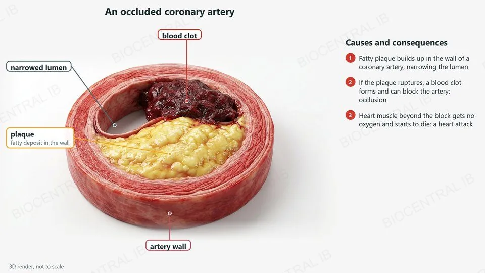
A 3D render of an occluded coronary artery, labelled: a fatty plaque in the wall, a blood clot, the narrowed lumen and the artery wall
B3.2.6

When a coronary artery is blocked

The coronary arteries supply the heart muscle itself with oxygen and glucose.
  • Cause: fatty plaque builds up in the wall of a coronary artery (atherosclerosis), narrowing the lumen.
  • Risk factors include high blood cholesterol, high blood pressure, smoking, obesity and lack of exercise.
  • If a plaque ruptures, a blood clot forms and can block the artery completely: occlusion.
  • Consequence: heart muscle beyond the block gets no oxygen and starts to die, a heart attack.
RememberOcclusion stops aerobic respiration in the heart muscle it supplies.
B3.2.6 · NOS

Correlation is not causation

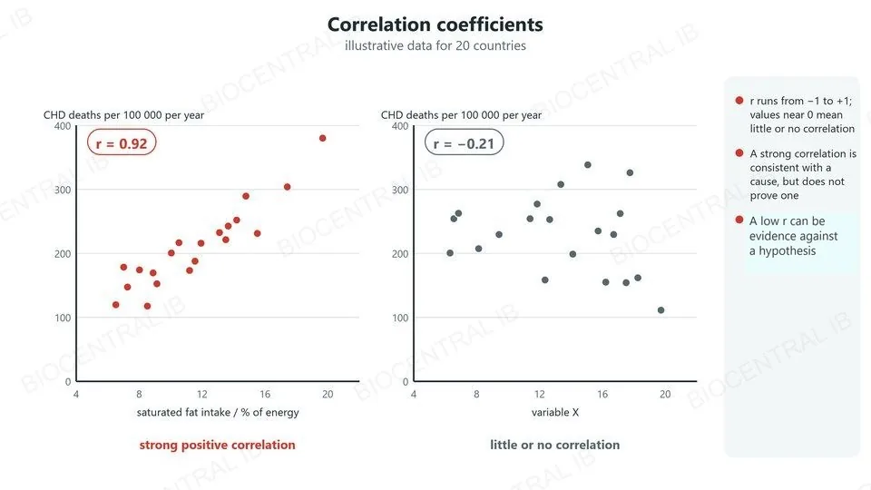
Epidemiological data on coronary heart disease are often analysed with correlation coefficients.
  • A correlation coefficient, r, quantifies a correlation: it runs from −1 to +1, and values near 0 mean little or no correlation.
  • A low r, or no correlation at all, can be evidence against a hypothesis.
  • Even a strong correlation, such as that between saturated fat intake and coronary heart disease, does not prove a causal link: another factor could affect both.
SkillEvaluate epidemiological data: strength of correlation, sample, and other factors.
Two scatter graphs of illustrative data: saturated fat intake against coronary heart disease deaths with r = 0.92, and an unrelated variable with r = −0.21
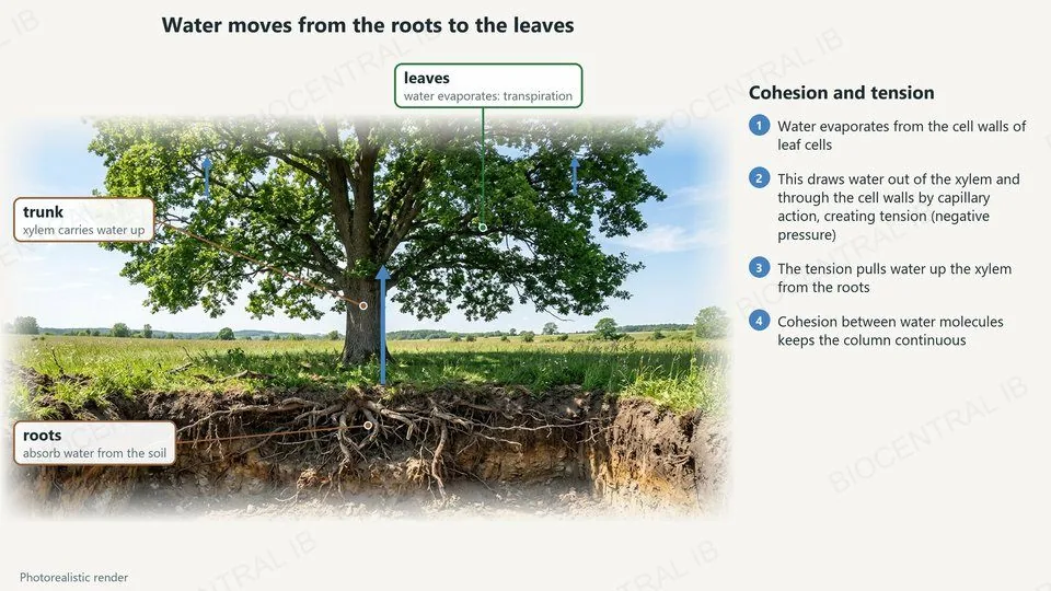
A photorealistic render of an oak tree with its roots exposed, labelled: leaves where water evaporates, the trunk where xylem carries water up, and roots that absorb water
B3.2.7

Water moves from roots to leaves

Water is carried up a plant in the xylem during transpiration, with no pump.
  • Water is lost by transpiration from the cell walls of leaf cells.
  • This draws water out of the xylem in the leaf, which pulls up water from the stem and the roots.
  • The roots absorb water from the soil to replace it.
CompareThe heart pushes blood; a transpiring leaf pulls water.
B3.2.7

Cohesion and tension

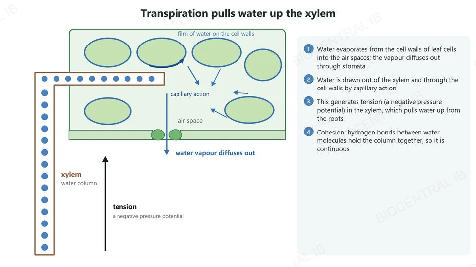
Transpiration generates tension that pulls water up the xylem.
  • Loss of water from the cell walls of leaf cells draws water out of the xylem vessels and through the cell walls by capillary action.
  • This generates tension, a negative pressure potential, in the xylem. The tension draws water up from the roots.
  • Cohesion: hydrogen bonds between water molecules hold them together, so the water column stays continuous.
LinkCohesion comes from hydrogen bonding between water molecules (A1.1).
Water evaporates from the wet cell walls of leaf cells into an air space and leaves through a stoma; water is drawn out of a xylem vessel, whose water column is under tension
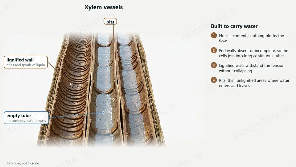
A 3D render of xylem vessels, labelled: lignified walls with rings and spirals of lignin, empty tubes with no end walls, and pits
B3.2.8

Xylem vessels: pipes under tension

Xylem vessels are adapted for the transport of water.
  • No cell contents and incomplete or absent end walls: the cells join into long, open tubes, so flow is unimpeded.
  • Lignified walls: lignin, in rings and spirals, makes the walls rigid enough to withstand the tension without collapsing.
  • Pits: thin, unlignified areas of the wall where water enters and leaves the vessels.
RememberXylem vessels are dead: the water moves passively.
B3.2.9 · Skill

A dicot stem in section

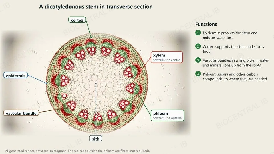
A transverse section of a dicotyledonous stem shows its tissues in fixed positions.
  • Epidermis: the outer layer, which protects the stem and reduces water loss.
  • Cortex: between the epidermis and the vascular bundles; it supports the stem and stores food.
  • Vascular bundles in a ring: xylem towards the centre carries water and mineral ions; phloem towards the outside carries sugars.
  • Pith: the central tissue.
RememberStem: a ring of bundles, xylem inside, phloem outside.
An AI-generated render, not a real micrograph, of a stained dicotyledonous stem section, labelled: epidermis, cortex, a ring of vascular bundles with xylem towards the centre and phloem towards the outside, and pith
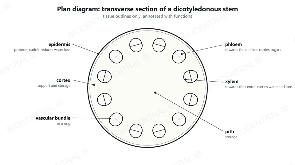
A plan diagram of a dicotyledonous stem: epidermis, cortex, a ring of vascular bundles with phloem outside and xylem inside, and pith, each annotated with its function
B3.2.9 · Skill

Plan diagram of a stem

A plan diagram shows the distribution of tissues, drawn from a micrograph.
  • Draw the outline of each tissue as a clear line; do not draw individual cells or use shading.
  • Show the relative positions of the epidermis, cortex, vascular bundles, xylem and phloem.
  • Annotate each structure with its main function.
SkillLabel lines should touch the tissue and never cross each other.
B3.2.10 · Skill

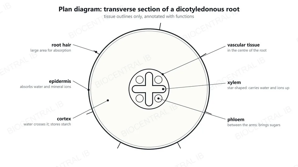
A dicot root in section

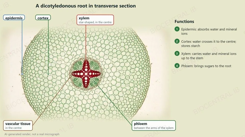
A transverse section of a dicotyledonous root shows a different arrangement from the stem.
  • Epidermis: the outer layer, with root hairs that absorb water and mineral ions.
  • Cortex: a wide layer that water crosses on its way to the centre; it stores starch.
  • Vascular tissue in the centre: xylem in a star or cross shape, and phloem in groups between the arms of the xylem.
CompareRoot: xylem in the centre. Stem: xylem in a ring of bundles.
An AI-generated render, not a real micrograph, of a stained dicotyledonous root section, labelled: epidermis, cortex, a central star of xylem with phloem between its arms
A plan diagram of a dicotyledonous root: root hairs, epidermis, cortex, central vascular tissue with a star of xylem and phloem between the arms, annotated with functions
B3.2.10 · Skill

Drawing a root section

The tissues of a root section can be drawn from a micrograph in the same way as a stem.
  • Draw the epidermis, the cortex and the central vascular tissue as outlines.
  • Draw the xylem as a star and the phloem as separate groups between its arms.
  • Annotate each tissue with its function: absorption, transport of water, transport of sugars.
SkillDraw what is in the micrograph, not a textbook picture.
Quick check

A micrograph shows a vessel with a thin wall around a wide lumen. Which vessel is it, and why?

an artery: it carries blood at high pressure
a vein: it carries blood at low pressure
a capillary: its wall is one cell thick
an arteriole: it controls the flow of blood
Correct answer: a vein. Blood in veins is at low pressure, so a thin wall is enough and the wide lumen lets blood flow with little resistance. An artery has a thick wall around a narrow lumen.
Part two · HL

Tissue fluid, the heart and phloem

B3.2.11 – B3.2.18 · HL only
B3.2.11 · HL

Tissue fluid: out and back in

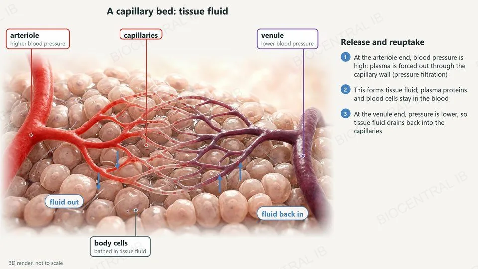
Tissue fluid is formed by pressure filtration of plasma in capillaries.
  • Blood from arterioles arrives at higher pressure, which forces plasma out through the capillary wall: pressure filtration.
  • Plasma proteins and blood cells are too large to leave, so they stay in the blood.
  • At the venule end the pressure is lower, so tissue fluid drains back into the capillaries.
RememberHigh pressure pushes fluid out; lower pressure lets it drain back.
A 3D render of a capillary bed between an arteriole at higher pressure and a venule at lower pressure; fluid leaves the capillaries near the arteriole and drains back near the venule
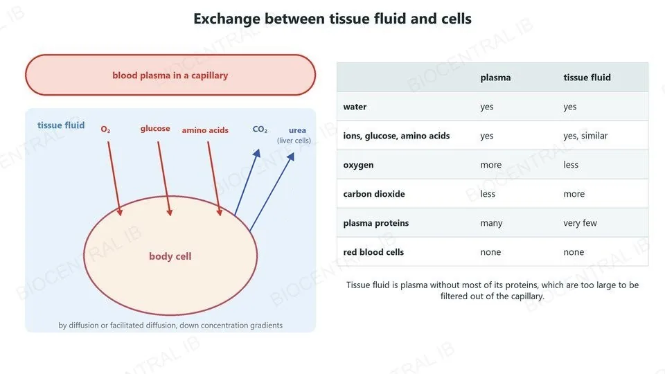
Oxygen, glucose and amino acids diffuse from tissue fluid into a body cell, and carbon dioxide and urea diffuse out; a table compares plasma with tissue fluid
B3.2.12 · HL

Exchange with the cells

Cells exchange substances with the tissue fluid that bathes them, not directly with the blood.
  • Oxygen, glucose and amino acids diffuse from the tissue fluid into the cells.
  • Carbon dioxide and other wastes such as urea diffuse from the cells into the tissue fluid.
  • Composition: tissue fluid is similar to plasma but has very few plasma proteins; near respiring cells it has less oxygen and more carbon dioxide.
RememberTissue fluid = plasma without most of its proteins.
B3.2.13 · HL

Excess fluid drains into lymph ducts

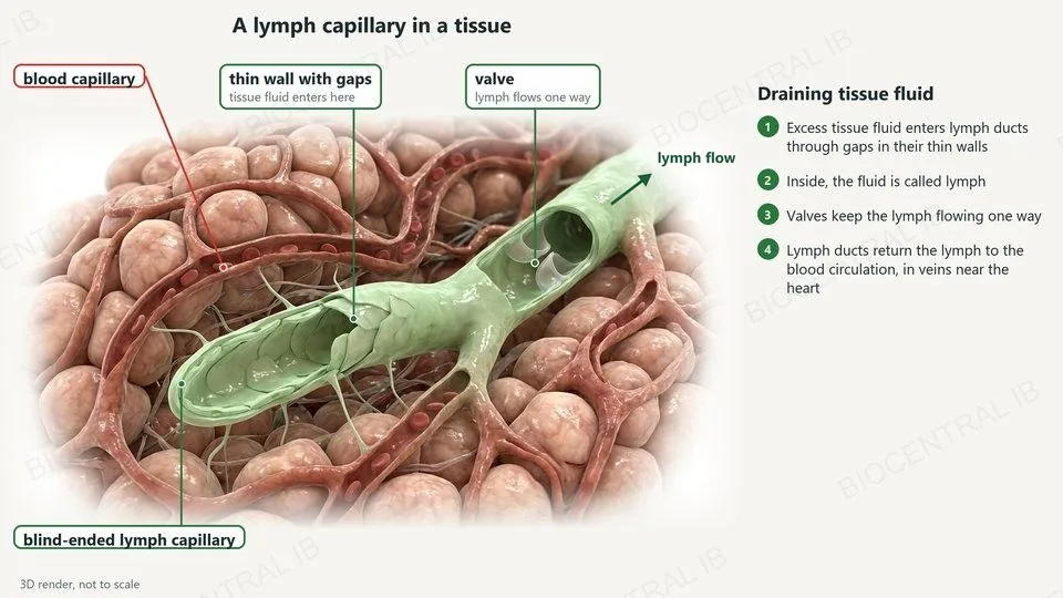
Not all tissue fluid returns to the capillaries; the excess drains into lymph ducts.
  • Lymph ducts have thin walls with gaps, where tissue fluid enters. Inside, the fluid is called lymph.
  • Valves in the lymph ducts keep the lymph flowing in one direction.
  • Lymph is returned to the blood circulation, in veins near the heart.
Why it mattersWithout drainage, tissue fluid would build up in the tissues.
A 3D render of a blind-ended lymph capillary among blood capillaries, labelled: a thin wall with gaps where tissue fluid enters, and a valve that keeps lymph flowing one way
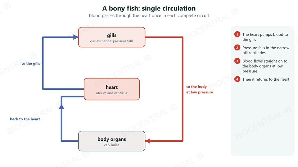
A circuit diagram of the single circulation of a bony fish: heart to gills, where pressure falls, then straight on to the body organs and back to the heart
B3.2.14 · HL

A fish: single circulation

In a bony fish, blood passes through the heart once in each complete circuit.
  • The heart pumps blood to the gills.
  • Blood pressure falls as it passes through the narrow gill capillaries.
  • It flows straight on to the body organs at low pressure, then returns to the heart.
RememberFish: heart → gills → body → heart.
B3.2.14 · HL

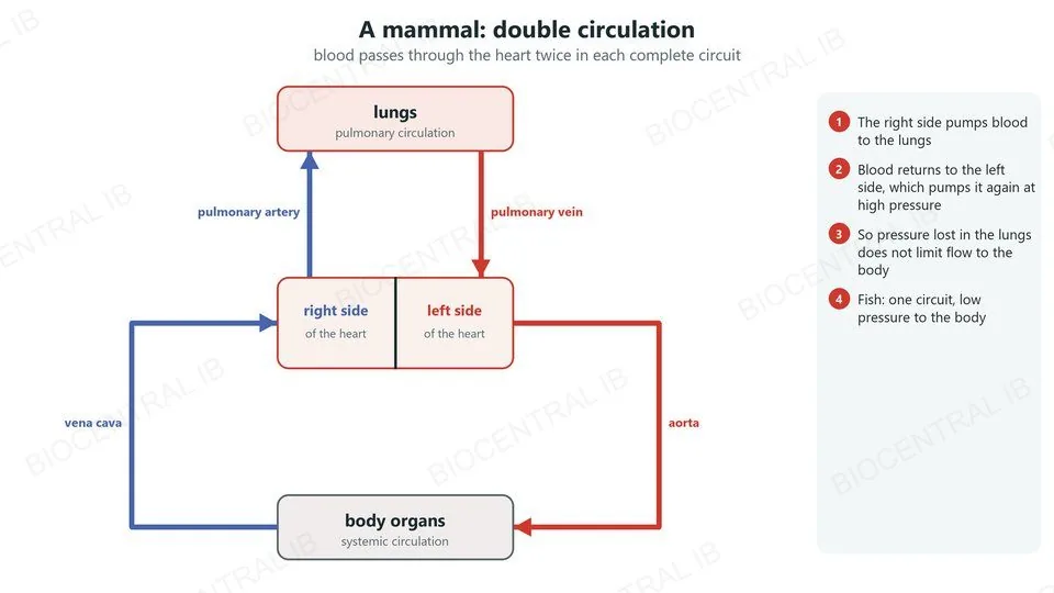
A mammal: double circulation

In a mammal, blood passes through the heart twice in each complete circuit.
  • The right side of the heart pumps blood to the lungs: the pulmonary circulation.
  • Blood returns to the left side, which pumps it again, at high pressure, to the body: the systemic circulation.
  • So the pressure lost in the lung capillaries does not limit the flow of blood to the body.
CompareMammal: heart → lungs → heart → body → heart.
A circuit diagram of the double circulation of a mammal: the right side of the heart pumps blood to the lungs and the left side pumps it to the body
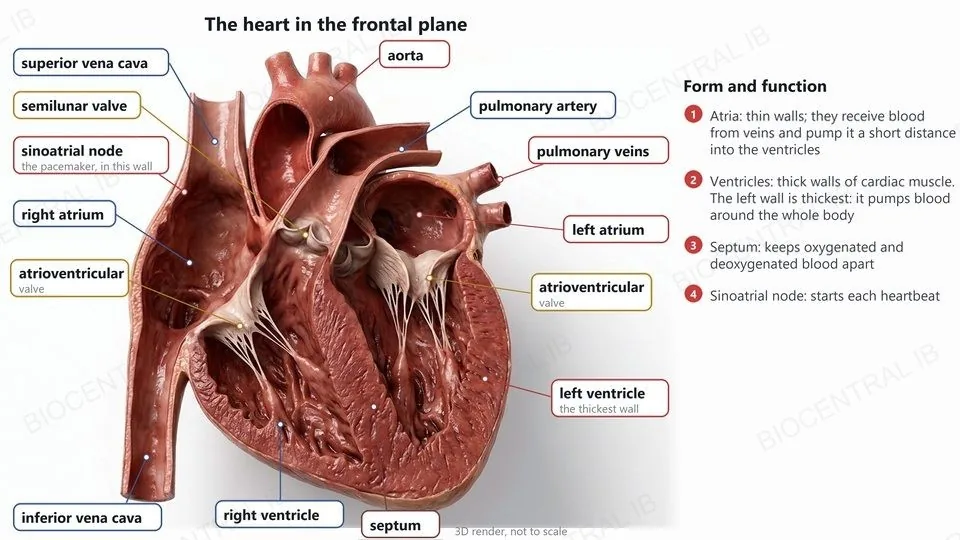
A 3D render of a heart cut in the frontal plane, labelled: sinoatrial node, atria, ventricles, septum, atrioventricular and semilunar valves, pulmonary veins, aorta, pulmonary artery and inferior vena cava
B3.2.15 · HL

The heart in the frontal plane

The structures of the mammalian heart are adapted to deliver pressurized blood to the arteries.
  • Atria: thin walls; they receive blood from veins and pump it a short distance into the ventricles.
  • Ventricles: thick walls of cardiac muscle; the left wall is thickest because it pumps blood around the whole body.
  • Septum: keeps oxygenated and deoxygenated blood apart.
  • Sinoatrial node (pacemaker): in the wall of the right atrium; it starts each heartbeat.
SkillIdentify these features on a diagram of the heart in the frontal plane.
B3.2.15 · HL

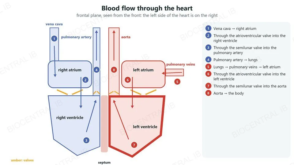
Tracing the flow of blood

Blood flows through the heart in one direction only, from named veins to named arteries.
  • Right side: vena cava → right atrium → atrioventricular valve → right ventricle → semilunar valve → pulmonary artery → lungs.
  • Left side: pulmonary veins → left atrium → atrioventricular valve → left ventricle → semilunar valve → aorta → body.
  • In the frontal plane, viewed from the front, the left side of the heart is on the right of the diagram.
SkillTrace the route from the vena cava to the aorta.
A diagram of the heart in the frontal plane with eight numbered steps of blood flow from the vena cava through the right side, the lungs and the left side to the aorta
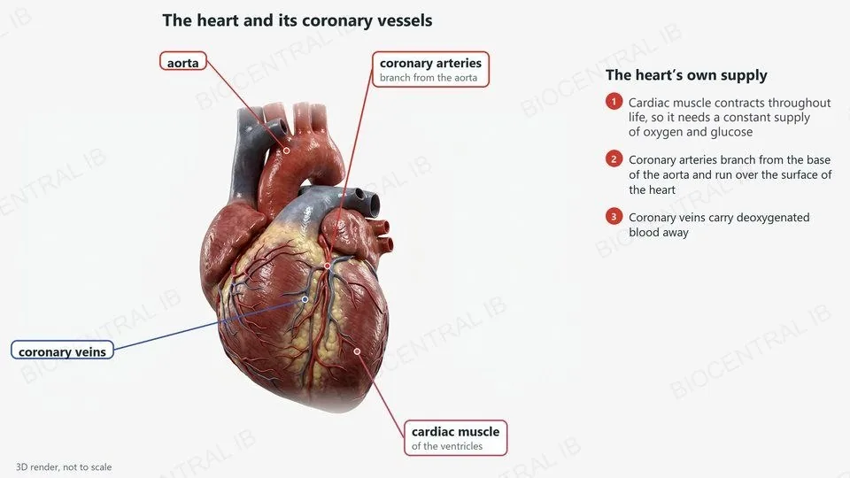
A 3D render of the front of a heart, labelled: aorta, coronary arteries branching from the aorta, coronary veins and the cardiac muscle of the ventricles
B3.2.15 · HL

The heart's own blood supply

Cardiac muscle contracts throughout life, so it needs a constant supply of oxygen and glucose for respiration.
  • Coronary arteries branch from the base of the aorta and run over the surface of the heart.
  • They carry oxygenated blood to the cardiac muscle of the atria and ventricles.
  • Coronary veins carry the deoxygenated blood away.
LinkA blocked coronary artery causes a heart attack (B3.2.6).
B3.2.15 · HL

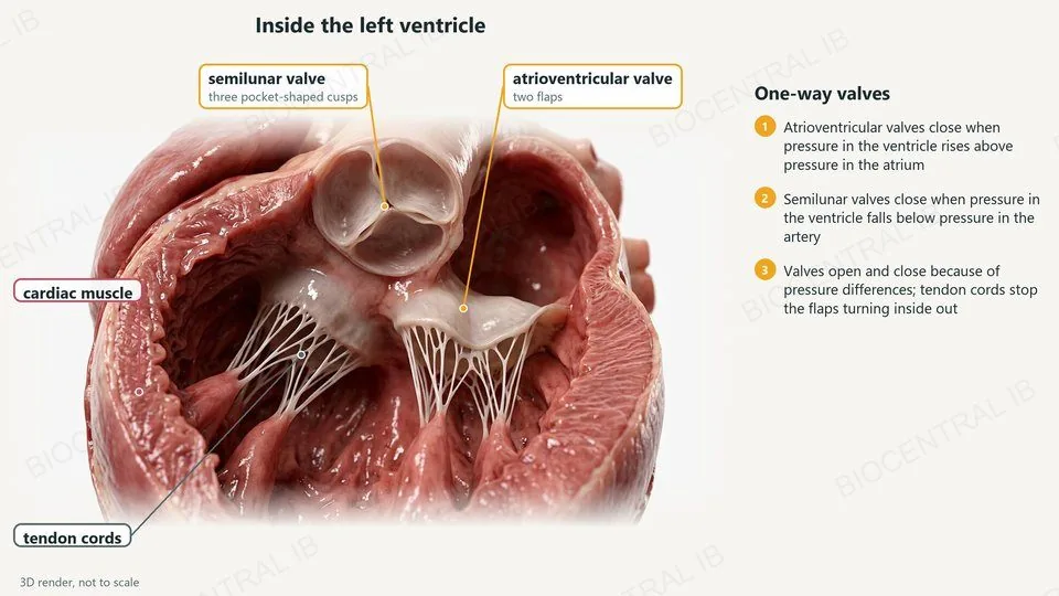
Valves: flow in one direction

Two sets of valves make sure blood flows only one way through the heart.
  • Atrioventricular valves, between the atria and the ventricles, close when pressure in the ventricle rises above pressure in the atrium.
  • Semilunar valves, at the bases of the aorta and pulmonary artery, close when pressure in the ventricle falls below pressure in the artery.
  • Valves open and close because of pressure differences; tendon cords stop the atrioventricular flaps turning inside out.
RememberValves are passive: pressure moves them.
A 3D render of the inside of a left ventricle, labelled: the semilunar valve with three cusps, the atrioventricular valve with two flaps, tendon cords and cardiac muscle
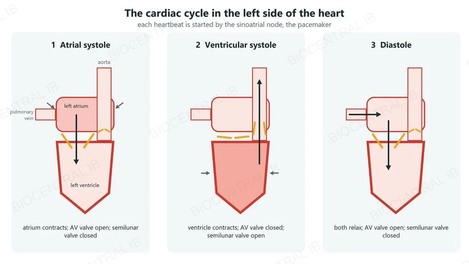
Three panels of the left side of the heart: atrial systole, ventricular systole and diastole, showing which chamber contracts and whether each valve is open or closed
B3.2.16 · HL

The cardiac cycle

Each heartbeat is started by the sinoatrial node, the pacemaker, and follows a fixed sequence of events.
  • Atrial systole: the atria contract, pushing more blood into the ventricles; atrioventricular valves open, semilunar valves closed.
  • Ventricular systole: the ventricles contract; the atrioventricular valves close, the semilunar valves open and blood is ejected into the arteries.
  • Diastole: the heart relaxes; the semilunar valves close, the atrioventricular valves open and the ventricles fill.
RememberSystole = contraction; diastole = relaxation.
B3.2.16 · HL · Skill

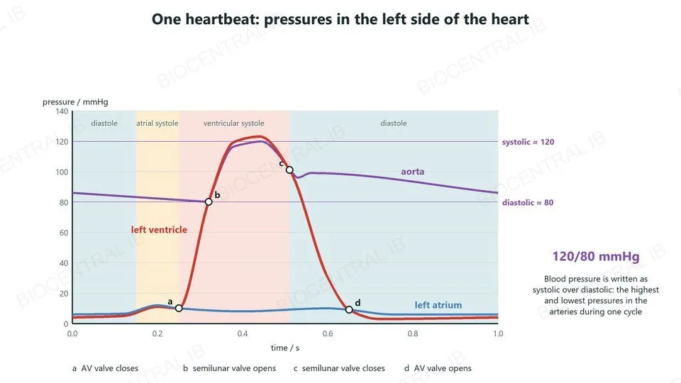
Reading blood pressure

Blood pressure is written as systolic over diastolic pressure, for example 120/80 mmHg.
  • Systolic: the highest pressure in the arteries, reached when the ventricles contract.
  • Diastolic: the lowest pressure, between beats, when the ventricles relax.
  • On a pressure graph, a valve opens or closes where two curves cross: the atrioventricular valve where ventricle and atrium cross, the semilunar valve where ventricle and aorta cross.
SkillRead systolic and diastolic pressures from data and graphs.
A graph of pressure in the left atrium, left ventricle and aorta over one heartbeat, with the moments the valves open and close and the systolic and diastolic pressures, 120/80 mmHg
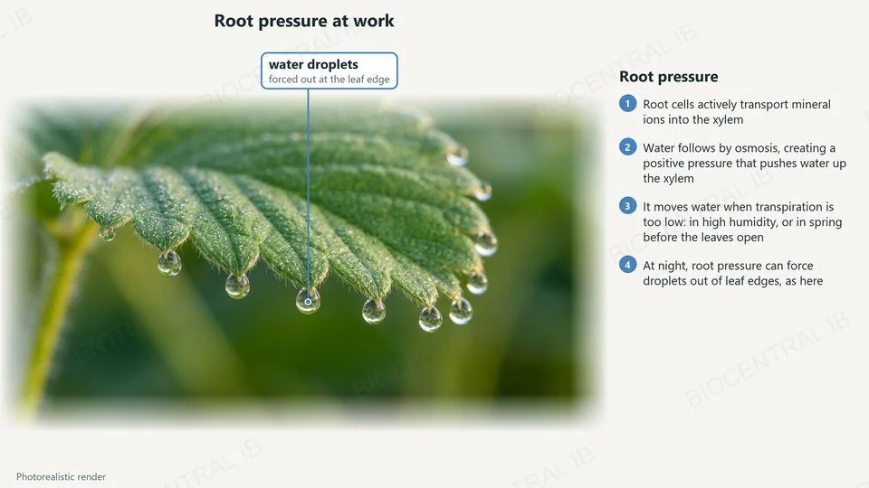
A photorealistic render of a strawberry leaf with water droplets forced out at the tips of its teeth by root pressure
B3.2.17 · HL

Root pressure

Root pressure is a positive pressure potential generated in the xylem of the roots.
  • Root cells use active transport to move mineral ions into the xylem; this needs ATP from respiration.
  • Water follows by osmosis, creating a positive pressure that pushes water up the xylem.
  • Root pressure moves water when transport by transpiration is insufficient, for example when high humidity prevents transpiration, or in spring before the leaves of deciduous plants open.
ExampleDroplets on leaf edges at dawn are forced out by root pressure.
B3.2.18 · HL

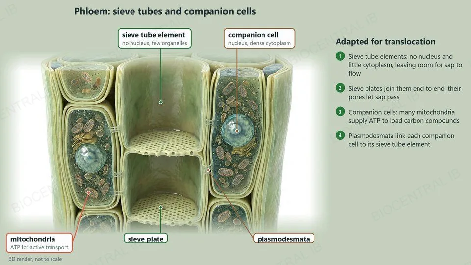
Sieve tubes and companion cells

Phloem sieve tubes and companion cells are adapted for the translocation of sap.
  • Sieve tube elements have no nucleus and reduced cytoplasm and organelles, leaving room for sap to flow.
  • Sieve plates join the elements end to end; their pores let sap pass.
  • Companion cells have many mitochondria, supplying ATP to load carbon compounds into the sieve tubes.
  • Plasmodesmata connect each companion cell to its sieve tube element.
RememberSieve tubes are living cells; xylem vessels are dead.
A 3D render of phloem, labelled: sieve tube elements with no nucleus, sieve plates, companion cells with a nucleus and many mitochondria, and plasmodesmata
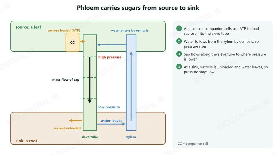
Phloem transport: companion cells load sucrose into a sieve tube at a leaf (source), water enters from the xylem, sap flows to a root (sink), where sucrose is unloaded and water leaves
B3.2.18 · HL

From source to sink

Sap flows in the phloem from sources, where carbon compounds are loaded, to sinks, where they are unloaded.
  • At a source, such as a leaf, companion cells load sucrose into the sieve tubes.
  • Water follows from the xylem by osmosis, so pressure rises.
  • Sap flows along the sieve tube to where the pressure is lower.
  • At a sink, such as a root or fruit, sucrose is unloaded and water leaves, so pressure stays low.
RememberHigh pressure at the source, low at the sink.
Quick check · HL

A student’s blood pressure is 124/78 mmHg. What does 78 mmHg represent?

the pressure when the ventricles contract
the pressure when the ventricles relax
the heart rate in beats per minute
the pressure in the left atrium
Correct answer: the pressure when the ventricles relax. 78 mmHg is the diastolic pressure, the lowest pressure in the arteries between beats. 124 mmHg, the systolic pressure, is reached when the ventricles contract.

Key vocabulary

Worth being able to define in a single sentence each

Capillary
a narrow vessel with a wall one cell thick, where exchange happens.
Fenestration
a pore in a capillary wall for rapid exchange.
Pulse
the surge of pressure in the arteries at each heartbeat.
Occlusion
complete blockage of an artery, for example by a clot.
Correlation coefficient
r, a number from −1 to +1 for the strength of a correlation.
Tension
negative pressure in the xylem created by transpiration.
Cohesion
hydrogen bonding that holds water molecules together.
Lignin
the polymer that makes xylem walls rigid.
Plan diagram
a drawing of tissue outlines, without individual cells.

Key vocabulary · HL

Additional higher level terms

Tissue fluid
fluid formed from plasma that bathes the cells.
Pressure filtration
plasma forced out of capillaries by blood pressure.
Lymph
excess tissue fluid in lymph ducts.
Double circulation
blood passes through the heart twice per circuit.
Sinoatrial node
the pacemaker that starts each heartbeat.
Systole / diastole
contraction / relaxation of the heart chambers.
Root pressure
positive pressure in xylem from active uptake of ions.
Companion cell
the cell that loads carbon compounds into a sieve tube.

Where this shows up again

B3.1 · Gas exchange
How do pressure differences contribute to the movement of materials in an organism?
A1.1 · Water
How do hydrogen bonds between water molecules let tension pull a continuous column of water up the xylem?
C1.2 · Cell respiration
How does respiration in sink tissues keep sucrose being unloaded from the phloem?
D2.3 · Water potential (HL)
How does water potential explain water moving from the xylem into sieve tubes at a source?

B3.2 Transport — one-page recap

Screenshot this slide to revise from

Blood vessels
  • Capillaries: branched, narrow, thin walls, some fenestrated.
  • Arteries: muscle + elastic tissue. Veins: thin, flexible walls, valves.
Pulse and coronary arteries
  • Pulse: radial or carotid; count and calculate beats per minute.
  • Plaque + clot → occlusion → heart attack. Correlation ≠ cause.
Xylem
  • Transpiration → tension; cohesion keeps the column continuous.
  • No contents, no end walls, lignified walls, pits.
Stem and root
  • Stem: ring of bundles, xylem inside, phloem outside.
  • Root: central star of xylem, phloem between the arms.
HL · Tissue fluid and circulation
  • Out at high pressure, back at lower pressure; excess drains into lymph.
  • Fish: single circulation. Mammal: double, repressurized.
HL · Heart, root pressure, phloem
  • SA node → atrial systole → ventricular systole → diastole; 120/80.
  • Root pressure: ions pumped, water follows. Phloem: source to sink.
Next step

Now test yourself on B3.2

Reading the slides is half the job. The other half is answering questions the way the exam asks them.

66 quiz questions7 data questions5 exam questionsmarkschemes included

Unlock every lesson with Pro — quizzes, data questions and exam papers. Two lessons are completely free to try: A1.1 Water and B1.1 Carbohydrates and lipids.

One-page summary, key terms and sample questions for B3.2 →

Pressure moves fluids.

A heart pushes blood, a leaf pulls water, and companion cells load sugar so that sap flows.
B3.2 Transport · BioCentral IB
Use ↓ ↑ or click to navigate
01 / 20