BioCentral IBBioCentral IB
IB Biology · Theme B · B3.1 · SL and HL

Gas exchange

How animals and plants exchange gases with their surroundings. Part one is for SL and HL; part two is HL only.
Guiding questions

How are multicellular organisms adapted to carry out gas exchange?

What are the similarities and differences in gas exchange between a flowering plant and a mammal?

Part one

Exchange surfaces in animals and plants

B3.1.1 – B3.1.10
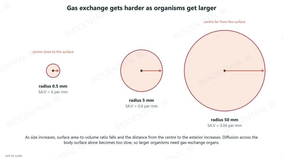
Three spheres of radius 0.5, 5 and 50 mm: as size increases, SA:V falls from 6 to 0.06 per mm and the centre gets further from the surface
B3.1.1

Gas exchange: a vital function

Every organism exchanges gases with its surroundings, for example taking in oxygen for respiration and releasing carbon dioxide.
  • Gases cross a gas-exchange surface by diffusion.
  • As an organism gets larger, its surface area-to-volume ratio falls.
  • The distance from its centre to its exterior also increases, so diffusion from the surface is too slow to supply the inner cells.
  • So larger organisms need gas-exchange surfaces and a system to transport gases.
ExampleAn amoeba exchanges gases across its plasma membrane; a mammal needs lungs.
B3.1.2

Properties of exchange surfaces

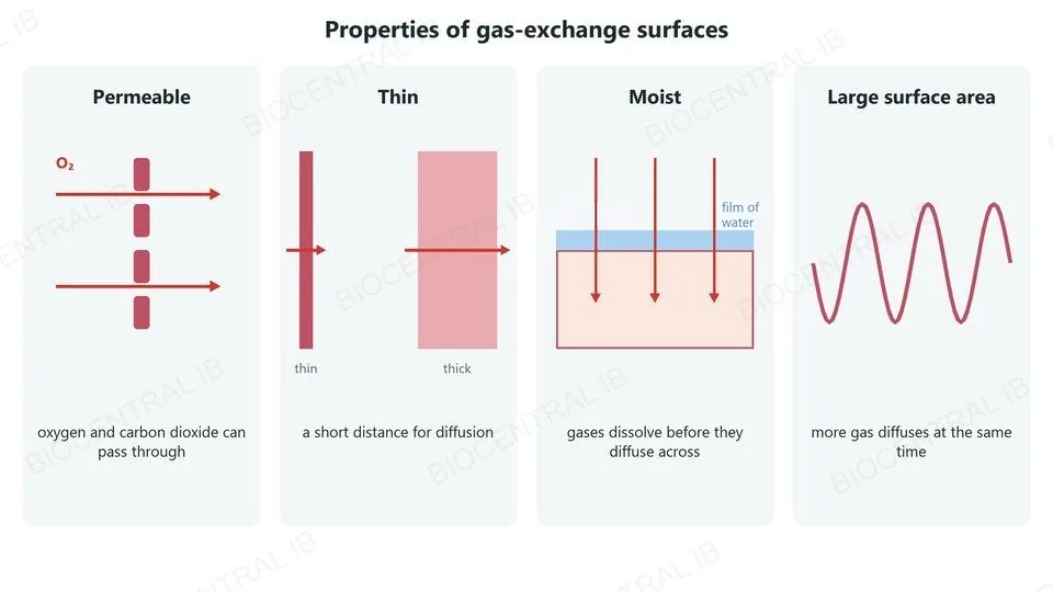
Gas-exchange surfaces in animals and plants share four properties.
  • Permeable: oxygen and carbon dioxide can pass through freely.
  • Thin tissue layer: the distance for diffusion is short.
  • Moist: gases dissolve in a film of water before they diffuse across.
  • Large surface area: more gas can diffuse at the same time.
ExamplesThe alveoli of lungs, the filaments of gills and the spongy mesophyll of leaves.
Four panels: a permeable surface lets oxygen through; a thin surface gives a short diffusion distance; a moist surface lets gases dissolve; a folded surface gives a large area
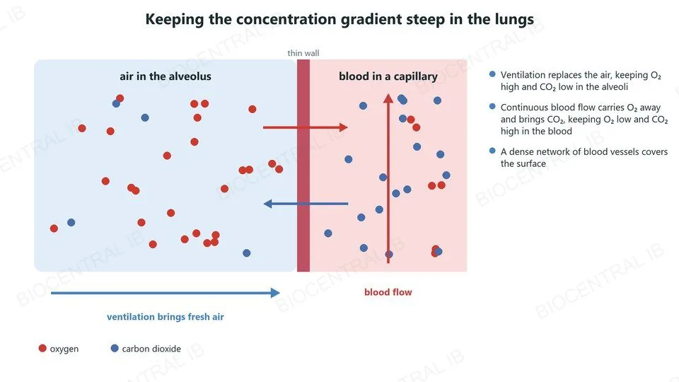
Air in an alveolus with much oxygen, a thin wall, and blood in a capillary with little oxygen; ventilation brings fresh air and blood flow carries oxygen away
B3.1.3

Keeping the gradient steep

Diffusion continues only while there is a concentration gradient, so animals keep the gradient steep at their exchange surfaces.
  • Dense networks of blood vessels cover the exchange surface.
  • Continuous blood flow carries oxygen away and brings carbon dioxide, so oxygen stays low and carbon dioxide high on the blood side.
  • Ventilation with air replaces the air in the lungs, so oxygen stays high and carbon dioxide low on the air side.
RememberBoth sides keep moving, so the gradient never runs down.
B3.1.3

Gills: ventilation with water

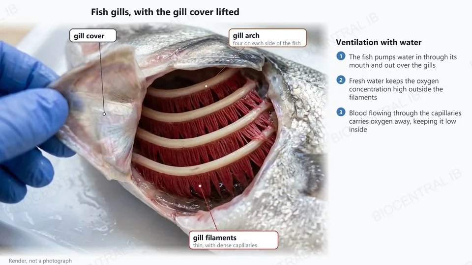
Fish keep the gradient steep at their gills by ventilating them with water.
  • Water is pumped in through the mouth and out over the gills, so water with more oxygen keeps arriving.
  • Each gill arch carries many thin filaments with a dense network of capillaries.
  • Blood flows continuously through the capillaries, carrying oxygen away from the gills and keeping its concentration low inside.
CompareLungs are ventilated with air; gills are ventilated with water.
A photorealistic render of fish gills with the gill cover lifted, labelled: gill cover, gill arch and thin gill filaments with dense capillaries
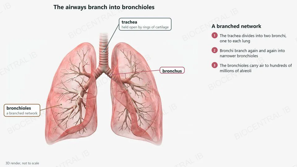
A 3D render of human lungs, labelled: the trachea held open by cartilage, a bronchus and the branched network of bronchioles
B3.1.4

A branched network of bronchioles

Mammalian lungs are adapted for gas exchange; air reaches the alveoli through a branched network of tubes.
  • The trachea, held open by rings of cartilage, divides into two bronchi, one to each lung.
  • The bronchi divide again and again, forming a branched network of bronchioles.
  • The bronchioles carry air to hundreds of millions of tiny air sacs, the alveoli, spread through the lungs.
Why it mattersBranching lets air reach a huge number of alveoli.
B3.1.4

Alveoli and capillary beds

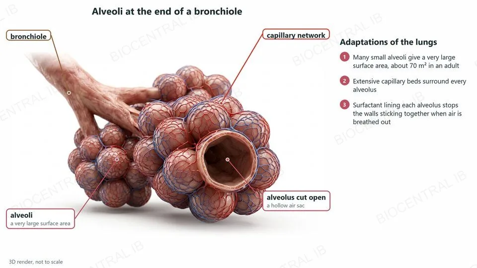
The alveoli give the lungs a very large surface area for gas exchange.
  • High surface area: many small alveoli give about 70 m² in an adult, far more than one large air sac could.
  • Extensive capillary beds surround the alveoli, so a large volume of blood is close to the air.
  • Surfactant coats the moist inner surface. It lowers surface tension, so the walls of the alveoli do not stick together when air is breathed out.
LinkThe cells of the alveolus wall, type I and type II pneumocytes, are in B2.3 (HL).
A 3D render of alveoli at the end of a bronchiole, labelled: bronchiole, alveoli, the capillary network and an alveolus cut open to show it is hollow
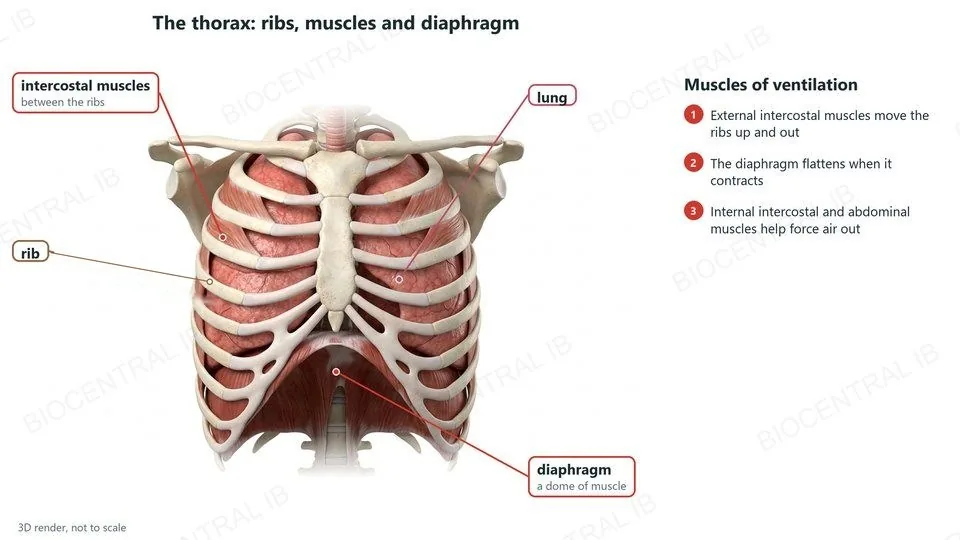
A 3D render of the thorax, labelled: intercostal muscles between the ribs, a rib, a lung and the dome-shaped diaphragm
B3.1.5

The muscles of ventilation

Ventilation moves air in and out of the lungs by changing the volume of the thorax.
  • Breathing in: the external intercostal muscles contract, moving the ribs up and out, and the diaphragm contracts and flattens.
  • The volume of the thorax increases, so the pressure in the lungs falls below atmospheric pressure and air flows in.
  • The muscles work in antagonistic pairs: the external and internal intercostal muscles, and the diaphragm and the abdominal muscles.
RememberMuscles change the volume; the change in pressure moves the air.
B3.1.5

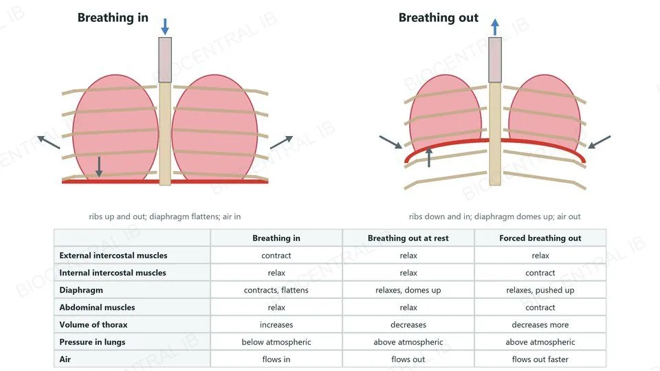
Breathing in and breathing out

Breathing out reverses the changes of breathing in.
  • At rest: the external intercostals and the diaphragm relax; the ribs move down and in and the diaphragm domes up.
  • Forced breathing out: the internal intercostal muscles contract, pulling the ribs down and in.
  • The abdominal muscles contract, pushing the abdominal organs and the diaphragm up.
  • The volume of the thorax decreases, so the pressure in the lungs rises above atmospheric pressure and air flows out.
RememberBreathing out at rest needs no muscle to contract.
Breathing in and breathing out side by side, with a table: external intercostals contract or relax, internal intercostals and abdominal muscles contract in forced breathing out, the diaphragm flattens or domes up, thorax volume and lung pressure change, and air flows in or out
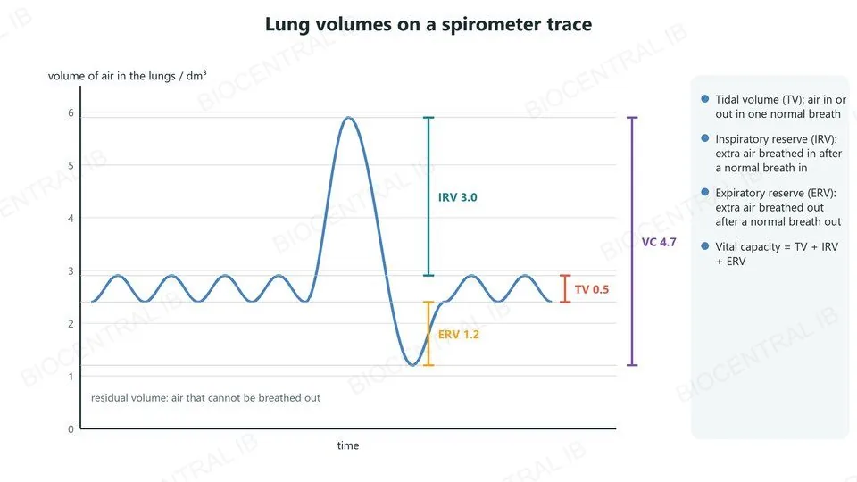
A spirometer trace of normal breaths, one deepest breath in and one fullest breath out, marked with tidal volume 0.5, inspiratory reserve 3.0, expiratory reserve 1.2 and vital capacity 4.7 cubic decimetres
B3.1.6 · Skill

Measuring lung volumes

Lung volumes can be measured with a spirometer, or by breathing out into an upturned bottle of water.
  • Tidal volume: the volume breathed in or out in one normal breath, about 0.5 dm³ at rest.
  • Inspiratory reserve: the extra volume that can be breathed in after a normal breath in. Expiratory reserve: the extra volume that can be breathed out after a normal breath out.
  • Vital capacity: the most air that can be breathed out after the deepest breath in; it equals tidal volume + inspiratory reserve + expiratory reserve.
SkillRepeat each measurement and use the mean.
B3.1.7

A leaf is adapted for gas exchange

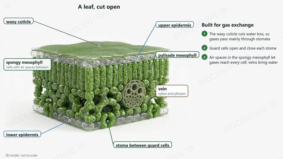
Leaves take in carbon dioxide and release oxygen for photosynthesis.
  • A waxy cuticle on the epidermis reduces water loss.
  • Stomata, pores in the epidermis, let gases in and out; guard cells open and close each one.
  • Air spaces in the spongy mesophyll let gases diffuse to and from every cell, giving a large surface area.
  • Veins bring water, which keeps the mesophyll cells moist, and carry products of photosynthesis away.
CompareLeaves and lungs both need a large, thin, moist surface.
A 3D cutaway render of a leaf, labelled: waxy cuticle, upper epidermis, palisade mesophyll, spongy mesophyll with air spaces, vein with xylem and phloem, lower epidermis and a stoma between guard cells
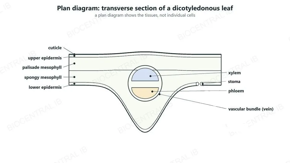
A plan diagram of a dicotyledonous leaf section labelled: cuticle, upper epidermis, palisade mesophyll, spongy mesophyll, lower epidermis, stoma, and a vascular bundle with xylem above and phloem below
B3.1.8 · Skill

Tissues in a leaf

A plan diagram shows the distribution of tissues in a transverse section of a dicotyledonous leaf, without drawing individual cells.
  • From the top: cuticle, upper epidermis, palisade mesophyll, spongy mesophyll and lower epidermis.
  • Most stomata are in the lower epidermis.
  • The vascular bundle (vein) runs through the mesophyll, with xylem towards the upper surface and phloem towards the lower surface.
SkillDraw each tissue boundary as one clear line and label every tissue.
B3.1.9

Transpiration

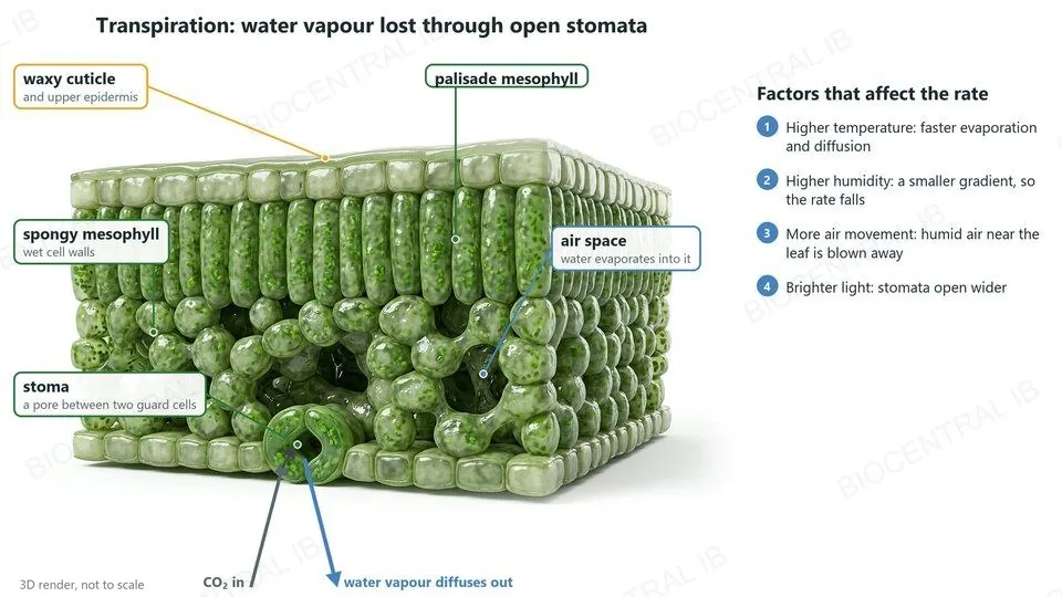
Transpiration is the loss of water vapour from leaves, a consequence of gas exchange.
  • Water evaporates from the moist walls of mesophyll cells into the air spaces, and water vapour diffuses out through open stomata.
  • Stomata must be open for carbon dioxide to enter, so some water is always lost.
  • The rate rises with higher temperature, lower humidity, more air movement and brighter light, which opens the stomata wider.
RememberThe pores that let carbon dioxide in also let water vapour out.
A 3D cutaway render of a leaf: waxy cuticle and upper epidermis, palisade mesophyll, spongy mesophyll with wet cell walls and air spaces, and a stoma, a pore between two guard cells. Water evaporates into the air spaces and water vapour diffuses out through the stoma while carbon dioxide diffuses in. The rate rises with higher temperature, lower humidity, more air movement and brighter light
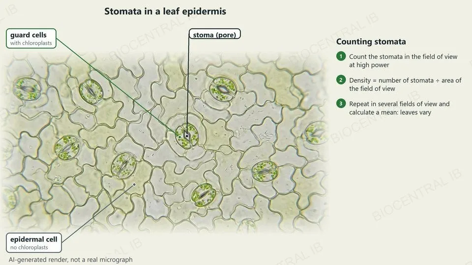
An AI-generated render, not a real micrograph, of leaf epidermis, labelled: guard cells with chloroplasts, a stoma, and an epidermal cell
B3.1.10 · Skill

Stomatal density

Stomatal density is the number of stomata per unit area of leaf surface.
  • Use a micrograph, or paint the leaf with clear nail varnish, peel off the cast and view it at high power.
  • Count the stomata in the field of view, then divide by the area of the field of view (πr²).
  • NOS: repeat the count in several fields of view and use the mean. Counts vary because biological material is variable, so replicates make the result more reliable.
Calculate14.8 stomata ÷ 0.1256 mm² ≈ 118 stomata per mm².
Quick check

Which change would increase the rate of transpiration from a leaf?

higher humidity
stiller air
lower temperature
more air movement
Correct answer: more air movement. Moving air carries away the humid air next to the leaf, keeping the water vapour gradient steep. Higher humidity, still air and lower temperature all reduce the rate.
Part two · HL

Haemoglobin and oxygen

B3.1.11 – B3.1.13 · HL only
B3.1.11 · HL

Haemoglobin

Haemoglobin in red blood cells transports oxygen.
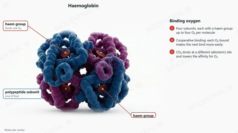
  • Each molecule has four polypeptide subunits, each with a haem group that can bind one oxygen molecule.
  • Cooperative binding: when one oxygen binds, the shape of the molecule changes, so the next oxygen binds more easily.
  • Allosteric binding of carbon dioxide: carbon dioxide binds at a site other than the haem groups, which lowers the affinity of haemoglobin for oxygen.
RememberOxygen binds at the haem groups; carbon dioxide binds elsewhere.
A molecular render of haemoglobin, labelled: four polypeptide subunits, each with a haem group that binds one oxygen molecule
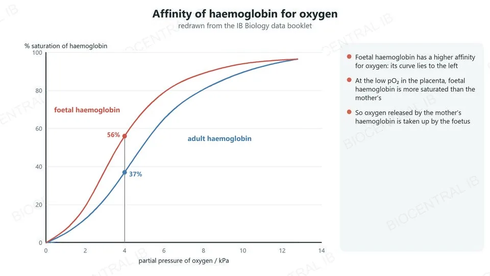
Oxygen dissociation curves of foetal and adult haemoglobin, redrawn from the data booklet: at 4 kilopascals foetal haemoglobin is about 56% saturated and adult about 37%
B3.1.11 · HL

Foetal and adult haemoglobin

Foetal haemoglobin has a higher affinity for oxygen than adult haemoglobin.
  • At any partial pressure of oxygen below the plateau, foetal haemoglobin is more saturated: its curve lies to the left.
  • In the placenta the partial pressure of oxygen is low, so the mother’s haemoglobin releases oxygen.
  • Foetal haemoglobin binds this oxygen, so oxygen passes from the mother’s blood to the foetus.
Data bookletThis graph is in the data booklet; it is redrawn here.
B3.1.12 · HL

The Bohr shift

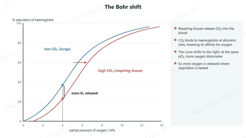
An increase in carbon dioxide causes haemoglobin to release more oxygen: the Bohr shift.
  • Respiring tissues release carbon dioxide into the blood.
  • Carbon dioxide binds to haemoglobin at allosteric sites and lowers its affinity for oxygen, so the dissociation curve shifts to the right.
  • At the same partial pressure of oxygen, more oxygen dissociates, so tissues that are respiring faster receive more oxygen.
Why it mattersOxygen is delivered where it is needed most.
Oxygen dissociation curves at low and high carbon dioxide: the high carbon dioxide curve lies to the right, so more oxygen is released at the same partial pressure
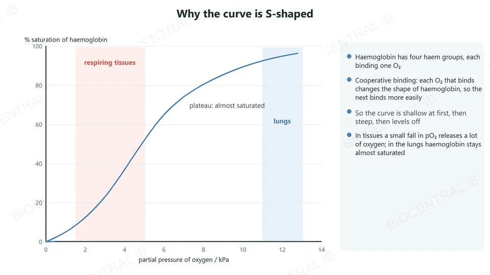
An S-shaped oxygen dissociation curve with the steep region at the partial pressures of respiring tissues and the plateau at the partial pressures in the lungs
B3.1.13 · HL

Why the curve is S-shaped

An oxygen dissociation curve shows the affinity of haemoglobin for oxygen at different oxygen concentrations.
  • The curve is S-shaped because of cooperative binding: once one oxygen has bound, the others bind more easily.
  • In the lungs the partial pressure of oxygen is high, so haemoglobin becomes almost fully saturated.
  • In respiring tissues the partial pressure is low, on the steep part of the curve, so a small fall releases a lot of oxygen.
SkillRead the saturation from the curve at a given partial pressure.
Quick check · HL

Blood passes through two tissues at the same partial pressure of oxygen. One tissue is producing more carbon dioxide. What happens there?

haemoglobin holds on to more oxygen
haemoglobin releases more oxygen
haemoglobin binds carbon dioxide at its haem groups
nothing changes
Correct answer: haemoglobin releases more oxygen. Carbon dioxide binds at allosteric sites, not the haem groups, and lowers the affinity of haemoglobin for oxygen: the Bohr shift.

Key vocabulary

Worth being able to define in a single sentence each

Ventilation
moving air or water over a gas-exchange surface.
Alveolus
a tiny air sac in the lungs where gases are exchanged.
Bronchiole
a narrow branch of the airways that carries air to the alveoli.
Surfactant
a secretion that stops the alveolus walls sticking together.
Tidal volume
the volume of air breathed in or out in one normal breath.
Vital capacity
tidal volume + inspiratory reserve + expiratory reserve.
Stoma
a pore in the leaf epidermis, opened and closed by guard cells.
Transpiration
loss of water vapour from leaves.
Stomatal density
the number of stomata per unit area of leaf.

Key vocabulary · HL

Additional higher level terms

Haemoglobin
the protein in red blood cells that transports oxygen.
Haem group
the part of each subunit that binds one oxygen.
Cooperative binding
each oxygen bound makes the next bind more easily.
Allosteric site
a binding site other than the one for oxygen.
Affinity
how strongly haemoglobin binds oxygen.
Bohr shift
the fall in oxygen affinity caused by more carbon dioxide.

Where this shows up again

B3.2 · Transport
How do multicellular organisms solve the problem of access to materials for all their cells?
C1.2 · Cell respiration
What is the relationship between gas exchange and metabolic processes in cells?
C3.1 · Integration of body systems
How do chemoreceptors and the brainstem control the ventilation rate?
B2.3 · Cell specialization (HL)
How are type I and type II pneumocytes adapted to their roles in the alveolus?

B3.1 Gas exchange — one-page recap

Screenshot this slide to revise from

Why gas exchange is hard
  • Larger size: lower SA:V and longer distances.
  • Surfaces: permeable, thin, moist, large area.
Gradients
  • Dense blood vessels and continuous blood flow.
  • Ventilation with air (lungs) or water (gills).
Lungs
  • Branched bronchioles; many alveoli; capillary beds.
  • Surfactant stops the alveoli collapsing.
Ventilation and volumes
  • In: external intercostals and diaphragm contract. Forced out: internal intercostals and abdominal muscles.
  • VC = TV + IRV + ERV.
Leaves
  • Cuticle, epidermis, palisade, spongy mesophyll, stomata, veins.
  • Transpiration; stomatal density with replicate counts.
HL · Haemoglobin
  • Cooperative binding gives an S-shaped curve; foetal Hb has higher affinity.
  • Bohr shift: CO₂ binds allosterically, more O₂ released.
Next step

Now test yourself on B3.1

Reading the slides is half the job. The other half is answering questions the way the exam asks them.

43 quiz questions6 data questions5 exam questionsmarkschemes included

Unlock every lesson with Pro — quizzes, data questions and exam papers. Two lessons are completely free to try: A1.1 Water and B1.1 Carbohydrates and lipids.

One-page summary, key terms and sample questions for B3.1 →

More area, less distance.

Lungs, gills and leaves solve the same problem in different ways.
B3.1 Gas exchange · BioCentral IB
Use ↓ ↑ or click to navigate
01 / 20