BioCentral IBBioCentral IB
IB Biology · Theme B · B2.3 · SL and HL

Cell specialization

How one cell becomes many kinds of cell. Part one is for SL and HL; part two is HL only.
Guiding questions

What are the roles of stem cells in multicellular organisms?

How are differentiated cells adapted to their specialized functions?

Part one

From one cell to many kinds

B2.3.1 – B2.3.6
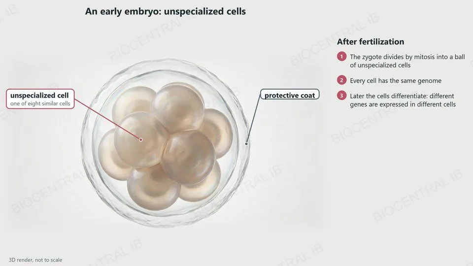
A 3D render of an early human embryo of eight similar, unspecialized cells inside a protective coat
B2.3.1

From zygote to specialized cells

After fertilization, the zygote divides to produce unspecialized cells, which later develop into specialized cells by differentiation.
  • The zygote divides again and again by mitosis, forming a ball of unspecialized embryonic cells.
  • All of these cells have the same genome: the same set of genes.
  • During differentiation, different genes are expressed in different cells, so each cell develops the structure that suits one function.
RememberSpecialized cells differ in which genes they use, not in the genes they have.
B2.3.1

Gradients and gene expression

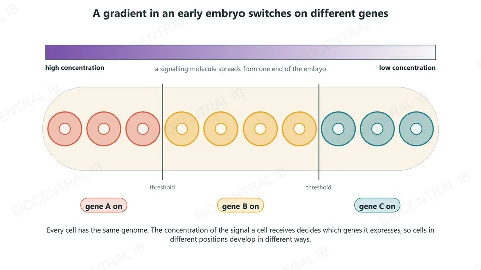
In an early-stage embryo, gradients of signalling molecules tell cells where they are.
  • A signalling molecule is released from one part of the embryo and diffuses away, so its concentration falls with distance.
  • Each cell detects the concentration where it is. Above or below certain thresholds, different genes are switched on.
  • So cells in different positions express different genes and develop along different pathways, even though their genomes are identical.
Why it mattersGradients like these lay out the body plan, for example from head to tail.
A signalling molecule spreads from one end of an embryo; cells at high, medium and low concentration switch on different genes
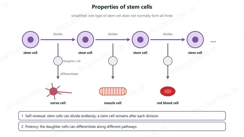
A stem cell divides again and again, and a stem cell remains each time; its other daughter cells differentiate into a nerve cell, a muscle cell or a red blood cell
B2.3.2

What makes a stem cell?

Stem cells have two properties together.
  • Self-renewal: they can divide endlessly, and a stem cell remains after each division.
  • Potency: their daughter cells can differentiate along different pathways, into different types of specialized cell.
  • Most specialized cells cannot do this: they divide rarely or never, and only make cells of their own type.
Why it mattersStem cells build the tissues of an embryo, and replace lost cells in adult tissues.
B2.3.3

Bone marrow: a stem cell niche

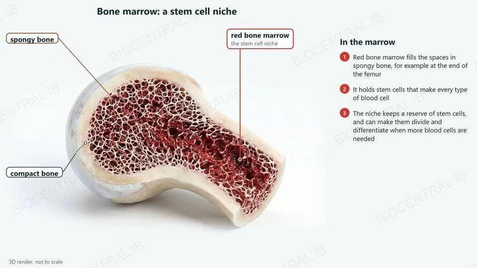
A stem cell niche is the place in the body where stem cells live; its conditions control what the stem cells do.
  • Red bone marrow, in the spaces of spongy bone, is the niche of the stem cells that produce blood cells.
  • The niche can maintain the stem cells, keeping a reserve that lasts for life.
  • It can also promote their proliferation and differentiation, so red blood cells, white blood cells and platelets are replaced as fast as they are lost.
ExampleBone marrow makes billions of new blood cells every day.
A 3D render of the end of a femur cut open, labelled: compact bone, spongy bone and red bone marrow, the stem cell niche
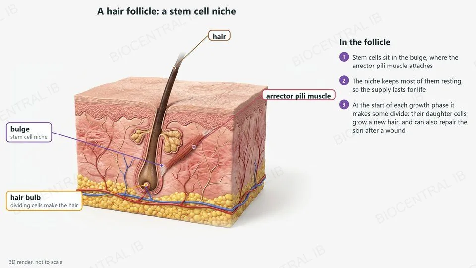
A 3D render of skin with a hair follicle, labelled: the hair, the arrector pili muscle, the bulge where stem cells live, and the hair bulb
B2.3.3

Hair follicles: a second niche

Each hair follicle has a stem cell niche in a region of its wall called the bulge, where the arrector pili muscle attaches.
  • Most of the time the niche keeps its stem cells resting, maintaining them for future hair growth.
  • When a new hair starts to grow, the niche promotes division: daughter cells move to the hair bulb, where cells proliferate and differentiate to form the hair.
  • After a wound, cells from the bulge also help to repair the skin.
RememberTwo niches to know: bone marrow and hair follicles.
B2.3.4

Totipotent, pluripotent, multipotent

Stem cells differ in the range of cell types they can become.
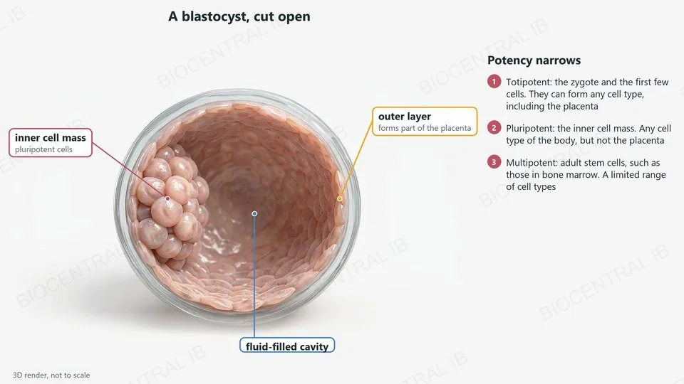
  • Totipotent: the zygote and the cells of the very early embryo. They can form any cell type, including the placenta.
  • Pluripotent: soon afterwards, the cells of the inner cell mass of the blastocyst. They can form any cell type of the body, but not the placenta.
  • Multipotent: stem cells in adult tissues such as bone marrow. They form only a limited range of related types, such as the types of blood cell.
RememberPotency narrows as development goes on.
A 3D render of a blastocyst cut open, labelled: the inner cell mass of pluripotent cells, the outer layer that forms part of the placenta, and the fluid-filled cavity
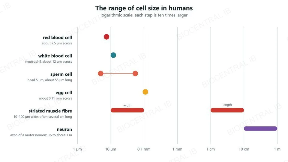
Human cell sizes on a logarithmic scale from 1 micrometre to 1 metre: red blood cell, white blood cell, sperm, egg, striated muscle fibre and neuron
B2.3.5

Cell size as specialization

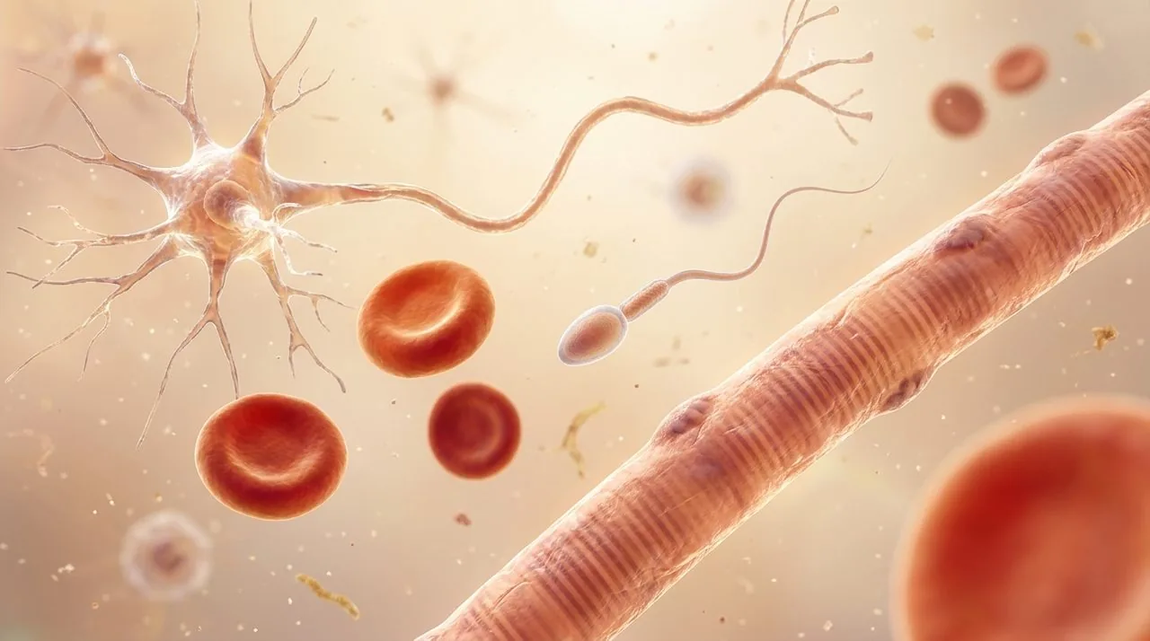
Cell size is an aspect of specialization: human cells range enormously in size.
  • Gametes: the egg is about 0.1 mm across and stores food; the sperm is tiny, with little cytoplasm, so it can swim.
  • Blood cells: red blood cells are small (about 7.5 µm) and squeeze through capillaries; white blood cells such as neutrophils are larger (about 12 µm).
  • Neurons: the axon of a motor neuron can be about 1 m long, from the spinal cord to the foot.
  • Striated muscle fibres: up to about 0.1 mm wide and often several centimetres long.
RememberEach cell has the size that suits its function.
B2.3.6

Surface area and volume

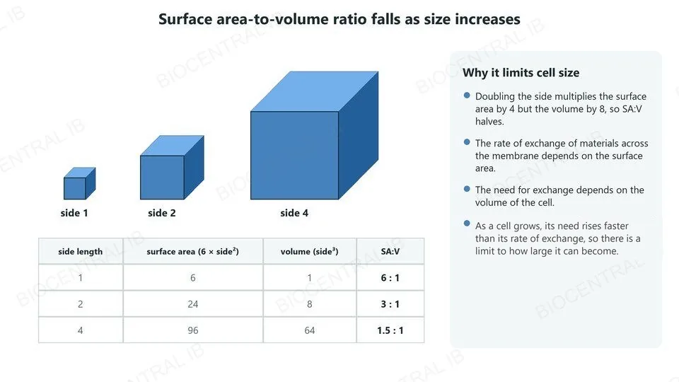
As a cell grows, its volume increases faster than its surface area.
  • For a cube, doubling the side multiplies the surface area by 4 (side²) but the volume by 8 (side³), so the surface area-to-volume ratio halves.
  • The rate of exchange of materials across the cell surface depends on its surface area.
  • The need for exchange, of oxygen, nutrients and waste, depends on the volume of the cell.
  • So beyond a certain size a cell cannot exchange materials fast enough: SA:V limits cell size.
CalculateA cube of side 2: surface area 24, volume 8, so SA:V = 3 : 1.
Cubes of side 1, 2 and 4 with their surface areas, volumes and surface area-to-volume ratios of 6, 3 and 1.5 to 1
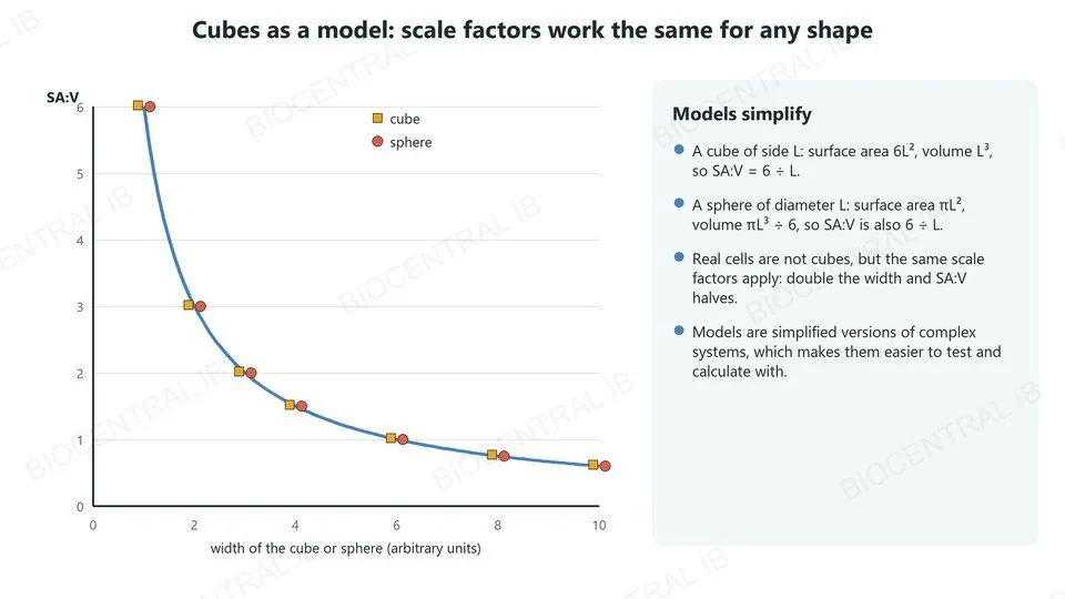
A graph of surface area-to-volume ratio against width: cubes and spheres fall on the same curve, 6 divided by width
B2.3.6 · Nature of science

Cubes as a model

Models are simplified versions of complex systems.
  • The surface area-to-volume relationship can be modelled using cubes of different side lengths.
  • Real cells are not cubes, and their shapes are more complex.
  • But scale factors work in the same way for any shape: a sphere of diameter L has the same SA:V as a cube of side L, 6 ÷ L.
  • So the simple model still correctly predicts that larger cells have a smaller SA:V.
Nature of scienceA good model leaves out detail but keeps what matters.
Quick check

A cell doubles in width but keeps the same shape. What happens to its surface area-to-volume ratio?

it doubles
it stays the same
it halves
it falls to one quarter
Correct answer: it halves. The surface area becomes 4 times larger (2²) and the volume 8 times larger (2³), so SA:V becomes 4 ÷ 8 = one half of what it was.
Part two · HL

Cells adapted to their functions

B2.3.7 – B2.3.10 · HL only
B2.3.7 · HL

Flattening: the erythrocyte

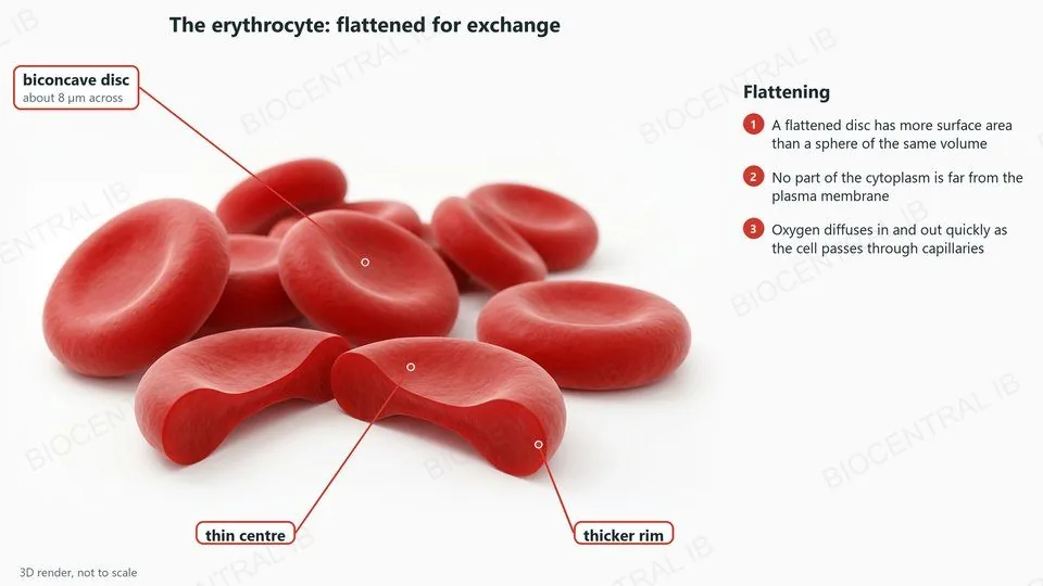
Cells can increase their surface area-to-volume ratio in three ways: flattening, microvilli and invagination.
  • Flattening: erythrocytes (red blood cells) are biconcave discs, thin in the centre and thicker at the rim.
  • A disc like this has about 40% more surface area than a sphere of the same volume, and no part of its cytoplasm is far from the membrane.
  • So oxygen diffuses in and out quickly while the cell passes through a capillary.
LinkType I pneumocytes are also flattened, coming up next.
A 3D render of red blood cells, labelled: a biconcave disc about 8 micrometres across, with a thin centre and a thicker rim
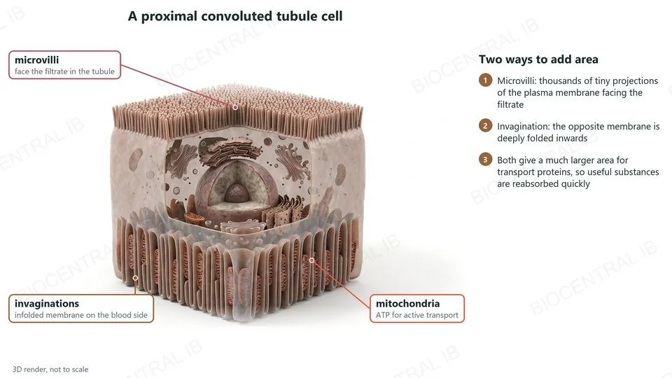
A 3D cutaway render of a proximal convoluted tubule cell, labelled: microvilli facing the filtrate, invaginations on the blood side, and mitochondria
B2.3.7 · HL

Microvilli and invagination

Cells of the proximal convoluted tubule in the nephron reabsorb useful substances from the filtrate.
  • Microvilli: the side facing the filtrate is covered in thousands of tiny projections of the plasma membrane.
  • Invagination: the plasma membrane on the side facing the blood is deeply folded inwards.
  • Both give a much larger area of membrane with more transport proteins, so glucose, ions and water are reabsorbed quickly.
  • Mitochondria between the infoldings supply ATP for active transport.
RememberMicrovilli project outwards; invaginations fold inwards.
B2.3.8 · HL

Type I pneumocytes

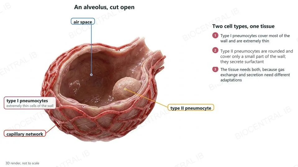
The alveolar epithelium is one tissue with two cell types, because gas exchange and secretion need different adaptations.
  • Type I pneumocytes cover most of the inner surface of each alveolus.
  • They are extremely thin, so the air in the alveolus and the blood in the capillaries are less than a micrometre apart.
  • This reduces the distance for diffusion of oxygen into the blood and of carbon dioxide out of it.
LinkGas exchange in the lungs is in B3.1.
A 3D render of an alveolus cut open, labelled: the air space, extremely thin type I pneumocytes, the capillary network and a type II pneumocyte
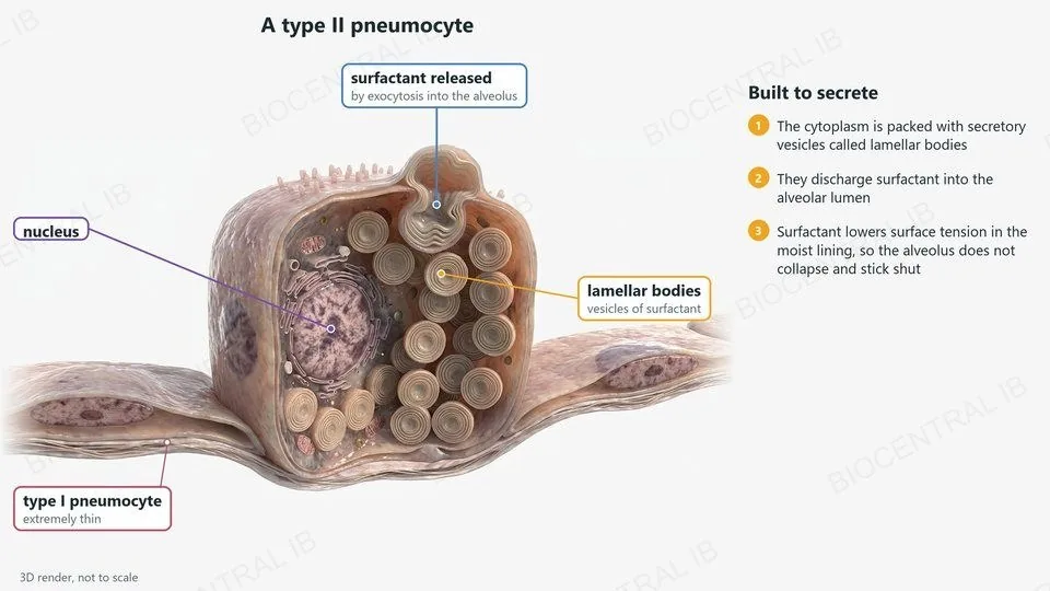
A 3D cutaway render of a type II pneumocyte, labelled: lamellar bodies, surfactant released by exocytosis, the nucleus, and a thin type I pneumocyte
B2.3.8 · HL

Type II pneumocytes

Type II pneumocytes are rounded cells scattered among the type I cells.
  • Their cytoplasm contains many secretory vesicles called lamellar bodies.
  • The lamellar bodies discharge surfactant into the alveolar lumen by exocytosis.
  • Surfactant lowers the surface tension of the film of moisture lining the alveolus, so the walls do not stick together and the alveolus does not collapse when air is breathed out.
CompareA cell thin enough for fast diffusion has no room for many vesicles, so two cell types are needed.
B2.3.9 · HL

Cardiac muscle cells

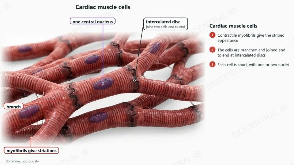
Cardiac muscle cells and striated muscle fibres both contain contractile myofibrils, but differ in other ways.
  • Both are packed with myofibrils, which shorten to cause contraction and give the striped (striated) appearance.
  • Cardiac muscle cells are short and branched, with one or two nuclei, and are joined end to end at intercalated discs.
  • Hypothesis: branching lets impulses spread in many directions through the wall of the heart, so it contracts as one unit.
RememberShort, branched, one or two nuclei: cardiac muscle.
A 3D render of cardiac muscle, labelled: branched cells joined at intercalated discs, one central nucleus and striations
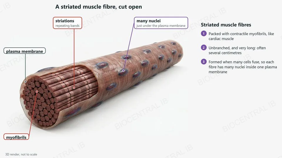
A 3D cutaway render of a striated muscle fibre, labelled: myofibrils, striations, many nuclei and the plasma membrane
B2.3.9 · HL

Is a muscle fibre a cell?

Striated muscle fibres are unbranched, very long and have many nuclei.
  • A fibre forms when many cells fuse during development, so it has many nuclei inside one plasma membrane.
  • Hypothesis: long fibres let a muscle shorten in one pull, and the many nuclei make enough proteins along the whole length.
  • Is a fibre a cell? It has one continuous plasma membrane and works as a unit. But it formed from many cells and has many nuclei, so it does not fit the usual idea of a cell.
DiscussThere is no single right answer: give reasons for your view.
B2.3.10 · HL

The sperm cell

The male gamete is adapted to swim to the egg and fertilize it.
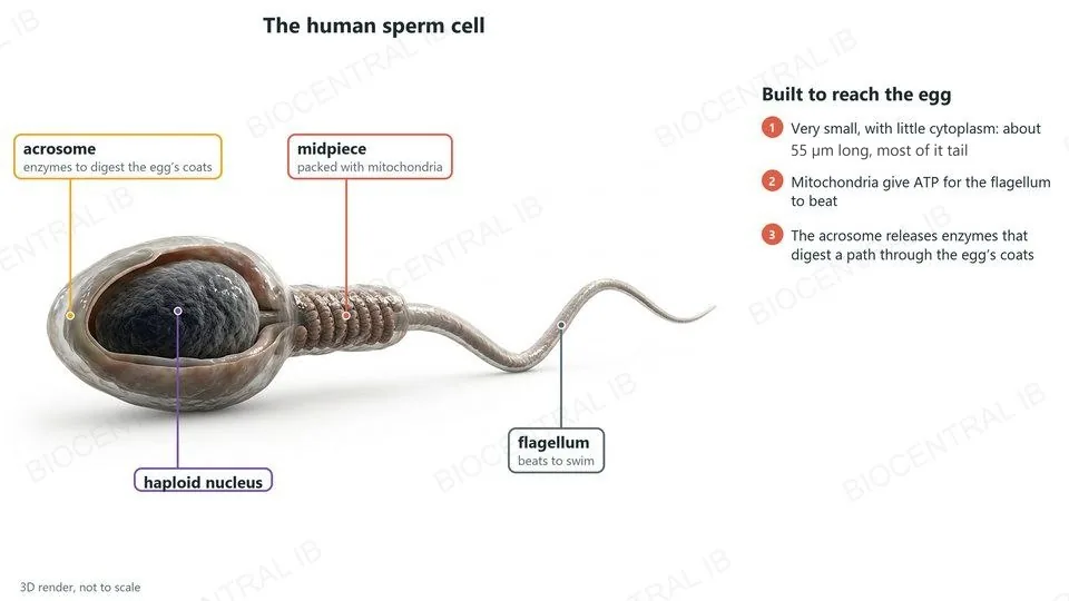
  • It is very small, with little cytoplasm; most of its length, about 55 µm, is the flagellum, which beats to swim.
  • The midpiece is packed with mitochondria, which produce the ATP the flagellum needs.
  • The head holds a haploid nucleus, capped by the acrosome: a vesicle of enzymes that digest a path through the coats around the egg.
LinkFertilization is in D3.1.
A 3D render of a human sperm cell, labelled: acrosome, haploid nucleus, midpiece with mitochondria and flagellum
A 3D render of a human egg cell cut open, labelled: follicle cells, zona pellucida, cytoplasm with food reserves, and sperm far smaller than the egg
B2.3.10 · HL

The egg cell

The female gamete is adapted to be fertilized and to start the embryo.
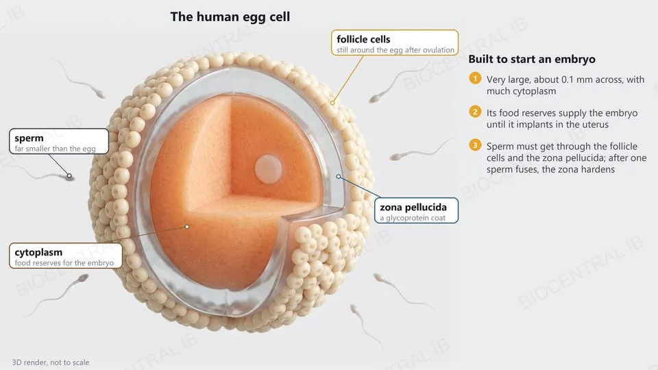
  • It is very large, about 0.1 mm across, with a lot of cytoplasm containing food reserves for the embryo until it implants.
  • It is surrounded by the zona pellucida, a glycoprotein coat, with a layer of follicle cells outside that.
  • When one sperm fuses with the egg, granules just inside its membrane release their contents and the zona pellucida hardens, so no other sperm can enter.
CompareThe sperm is small and moves; the egg is large and provides.
Quick check · HL

Why are type I pneumocytes extremely thin?

to secrete surfactant into the alveolus
to reduce the distance for diffusion of gases
to make room for many lamellar bodies
to increase the volume of the alveolus
Correct answer: to reduce the distance for diffusion of gases. Oxygen and carbon dioxide only have to cross a very short distance between the air and the blood. Surfactant is secreted from the lamellar bodies of type II pneumocytes.

Key vocabulary

Worth being able to define in a single sentence each

Differentiation
development of an unspecialized cell into a specialized cell by expressing particular genes.
Gradient
a change in the concentration of a signalling molecule across an embryo.
Stem cell
a cell that can divide endlessly and differentiate along different pathways.
Stem cell niche
the place where stem cells live, which maintains them or makes them divide and differentiate.
Totipotent
able to form any cell type, including the placenta.
Pluripotent
able to form any cell type of the body, but not the placenta.
Multipotent
able to form a limited range of related cell types.
SA:V
surface area divided by volume; it falls as a cell gets larger.
Model
a simplified version of a complex system, such as cubes for cells.

Key vocabulary · HL

Additional higher level terms

Microvilli
tiny projections of the plasma membrane that increase its area.
Invagination
inward folding of the plasma membrane.
Type I pneumocyte
an extremely thin alveolar cell, for diffusion of gases.
Type II pneumocyte
a rounded alveolar cell that secretes surfactant.
Lamellar body
a secretory vesicle of surfactant in a type II pneumocyte.
Surfactant
a secretion that lowers surface tension in the alveoli.
Myofibril
a contractile structure in muscle cells and fibres.
Intercalated disc
the junction joining cardiac muscle cells end to end.
Acrosome
a vesicle of enzymes on the head of a sperm.

Where this shows up again

D2.2 · Gene expression (HL)
How can cells with the same genome come to express different genes?
B3.1 · Gas exchange
Which adaptations of the alveoli make the lungs an efficient gas-exchange surface?
B3.3 · Muscle and motility (HL)
How do the myofibrils of a striated muscle fibre contract?
D3.1 · Reproduction
How do the structures of the sperm and the egg take part in fertilization?

B2.3 Cell specialization — one-page recap

Screenshot this slide to revise from

Differentiation
  • Zygote → unspecialized cells → specialized cells.
  • Same genome; gradients switch on different genes.
Stem cells and niches
  • Divide endlessly and differentiate along different pathways.
  • Niches maintain them or promote division: bone marrow, hair follicles.
Potency and size
  • Totipotent → pluripotent → multipotent.
  • Sizes: gametes, blood cells, neurons, muscle fibres.
SA:V
  • Exchange depends on area; need depends on volume.
  • SA:V falls as cells grow. NOS: cubes as a model.
HL · More surface
  • Flattening: erythrocytes. Microvilli and invagination: PCT cells.
  • Type I pneumocytes thin; type II secrete surfactant.
HL · Muscle and gametes
  • Cardiac: branched, 1–2 nuclei. Striated fibre: long, many nuclei.
  • Sperm: small, motile. Egg: large, food reserves.
Next step

Now test yourself on B2.3

Reading the slides is half the job. The other half is answering questions the way the exam asks them.

31 quiz questions6 data questions5 exam questionsmarkschemes included

Unlock every lesson with Pro — quizzes, data questions and exam papers. Two lessons are completely free to try: A1.1 Water and B1.1 Carbohydrates and lipids.

One-page summary, key terms and sample questions for B2.3 →

Same genes, different jobs.

Every specialized cell uses the same genome in its own way.
B2.3 Cell specialization · BioCentral IB
Use ↓ ↑ or click to navigate
01 / 20