BioCentral IBBioCentral IB
IB Biology · Theme B · B2.1 · SL and HL

Membranes and membrane transport

How lipids and proteins build a boundary that decides what enters and leaves a cell. Parts one to three are for SL and HL; part four is HL only.
Guiding questions

How do molecules of lipid and protein assemble into biological membranes?

What determines whether a substance can pass through a biological membrane?

Part one

The lipid bilayer and its proteins

B2.1.1 – B2.1.4
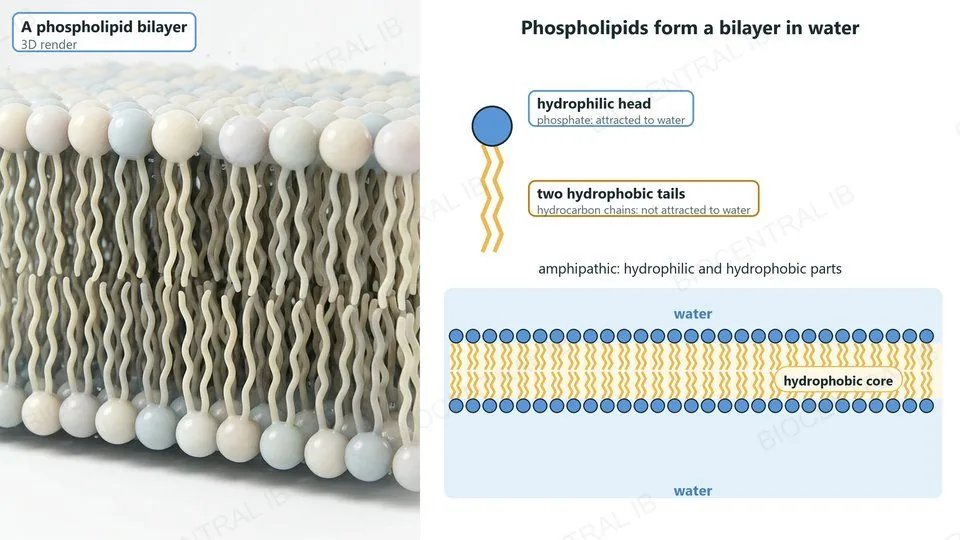
A 3D render of a phospholipid bilayer beside a diagram: one phospholipid with a hydrophilic phosphate head and two hydrophobic tails, and a bilayer in water with the heads facing the water on both sides and the tails forming a hydrophobic core
B2.1.1

Lipid bilayers

Lipid bilayers are the basis of cell membranes.
  • Phospholipids are amphipathic: each has a hydrophilic phosphate head and two hydrophobic hydrocarbon tails.
  • In water, phospholipids and other amphipathic lipids naturally form continuous, sheet-like bilayers.
  • The heads face the water on both sides, and the tails face each other in the middle, away from the water.
LinkThe structure of phospholipids is in B1.1.
B2.1.2

The bilayer is a barrier

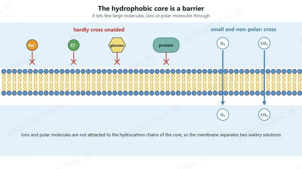
The core of the membrane is made of hydrophobic hydrocarbon chains.
  • This core has low permeability to large molecules, such as proteins, and to hydrophilic particles.
  • Hydrophilic particles include ions, such as Na⁺ and Cl⁻, and polar molecules, such as glucose.
  • So a membrane is an effective barrier between two aqueous solutions: the cytoplasm and the fluid outside the cell.
Why it mattersBecause the bilayer blocks most solutes, the cell can control what passes, using proteins.
A bilayer with Na⁺ and Cl⁻ ions, glucose and a protein unable to cross, while oxygen and carbon dioxide pass straight through
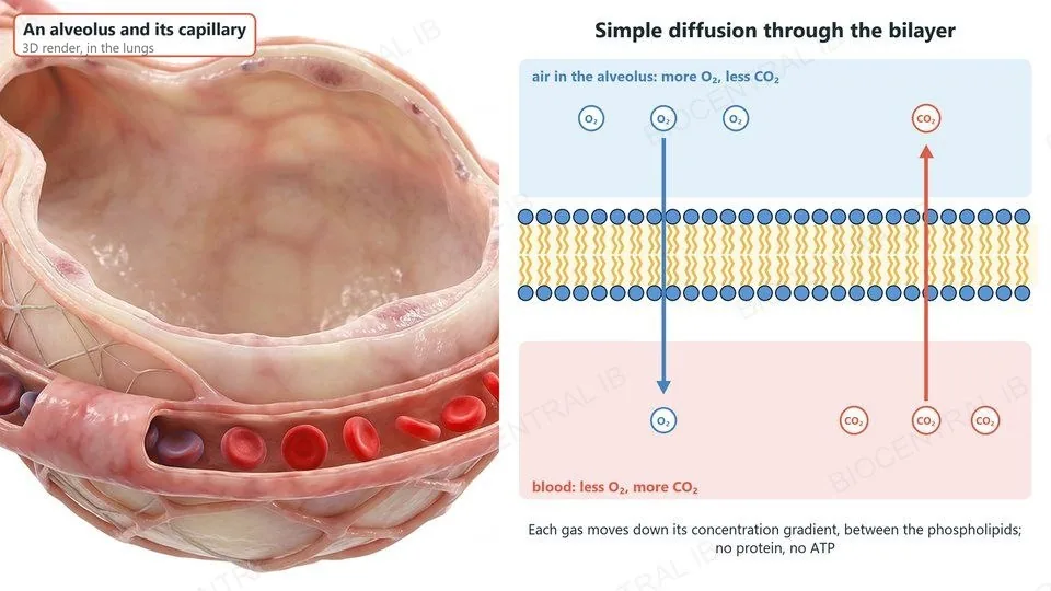
A cutaway alveolus with its capillary, beside a diagram of oxygen diffusing from the air in the alveolus through the bilayer into the blood, and carbon dioxide diffusing the other way, each down its concentration gradient
B2.1.3

Simple diffusion across membranes

Simple diffusion is the net movement of particles from a higher to a lower concentration, directly through the bilayer.
  • Oxygen and carbon dioxide are small and non-polar, so they pass between the phospholipids.
  • In the lungs, oxygen diffuses from the air in the alveoli into the blood, and carbon dioxide diffuses from the blood into the air.
  • Simple diffusion is passive: it needs no protein and no energy from ATP.
RememberDiffusion is always down a concentration gradient.
B2.1.4

Integral and peripheral proteins

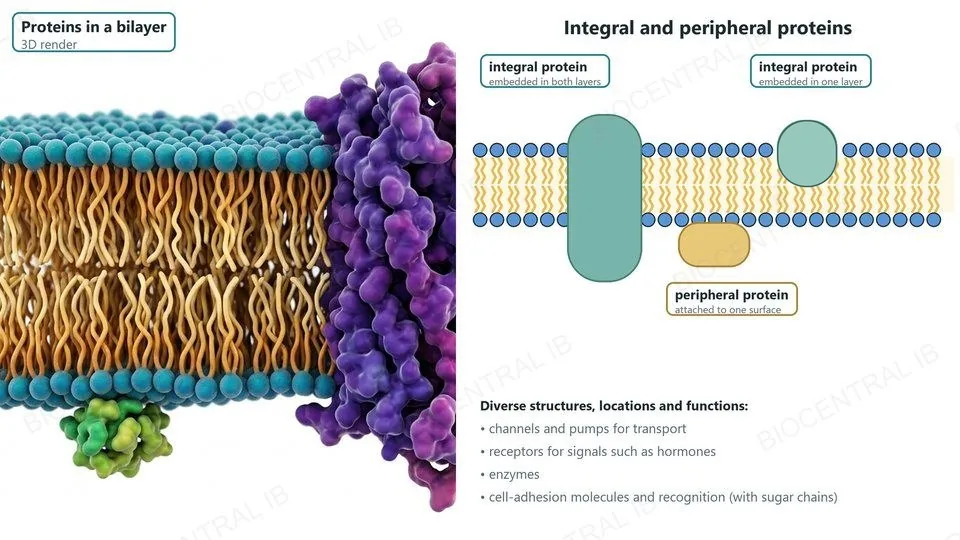
Membrane proteins have diverse structures, locations and functions.
  • Integral proteins are embedded in one or both of the lipid layers; many span the whole membrane.
  • Peripheral proteins are attached to one or other surface of the bilayer, without entering the core.
  • Membrane proteins act as channels and pumps, receptors for signals such as hormones, enzymes, and cell-adhesion molecules.
Why it mattersThe proteins in a membrane decide what it can do.
A 3D render of a bilayer with a protein spanning it and a small protein on the lower surface, beside a diagram of integral proteins embedded in both layers or one layer and a peripheral protein attached to the inner surface, with a list of functions
Part two

Moving across membranes

B2.1.5 – B2.1.8
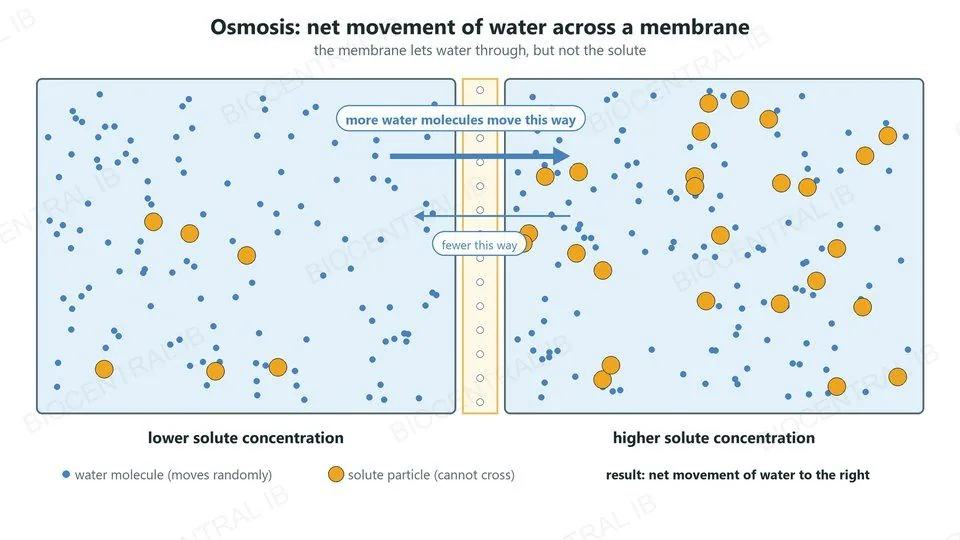
Two solutions separated by a membrane permeable to water but not to the solute. Water molecules move randomly both ways, but more move from the lower solute concentration to the higher solute concentration: a net movement of water
B2.1.5

Osmosis

Osmosis is the net movement of water across a membrane, from a lower to a higher solute concentration.
  • Water molecules move randomly and cross the membrane in both directions.
  • The membrane is impermeable to many solutes, so they stay on their own side.
  • Where the solute concentration is higher, fewer water molecules are free to move across, so more water moves towards that side than away from it.
ExampleA red blood cell in pure water takes in water, swells and bursts.
B2.1.5

Aquaporins

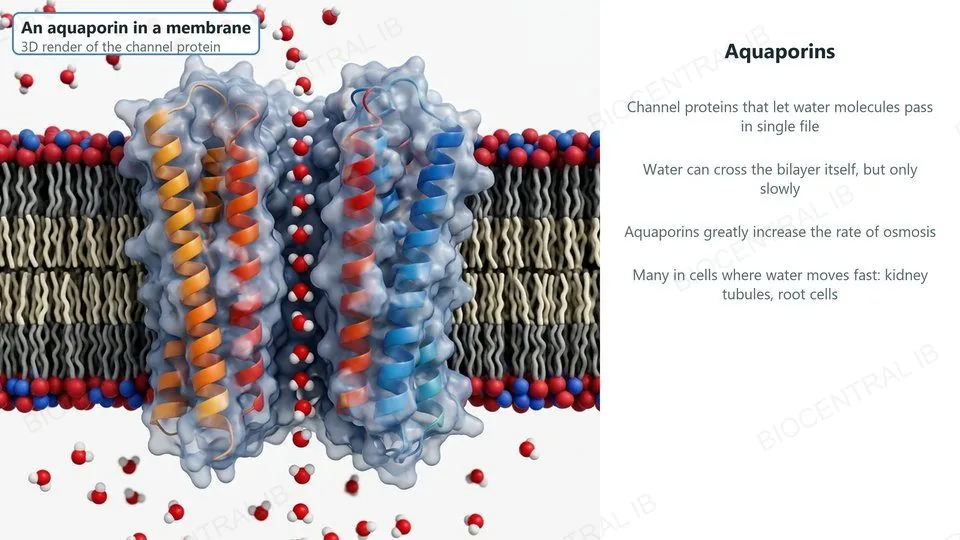
Aquaporins are channel proteins that let water molecules cross membranes rapidly.
  • Water can cross the bilayer itself, but only slowly, because water molecules are polar.
  • An aquaporin forms a narrow channel through which water molecules pass in single file.
  • Cells where water must move quickly, such as kidney tubule cells and root cells, have many aquaporins.
RememberAquaporins speed up osmosis; they do not pump water.
A 3D render of an aquaporin channel protein in a bilayer with water molecules passing through in single file, with notes on its role
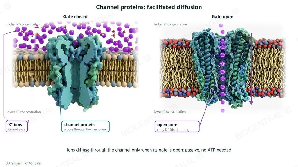
Two 3D renders of a potassium channel in a membrane. Gate closed: K⁺ ions on the higher-concentration side cannot pass. Gate open: K⁺ ions pass one at a time through the pore, down their concentration gradient; only K⁺ fits its lining. Passive, no ATP needed
B2.1.6

Channel proteins

Channel proteins allow facilitated diffusion: passive movement of particles through a protein, down a concentration gradient.
  • A channel forms a pore through the membrane with a particular diameter and lining.
  • Its structure lets only specific ions through, for example only K⁺ or only Na⁺.
  • Many channels have gates: the ions diffuse through when the channel is open, but not when it is closed.
Why it mattersChannels make the membrane selectively permeable.
B2.1.7

Pump proteins

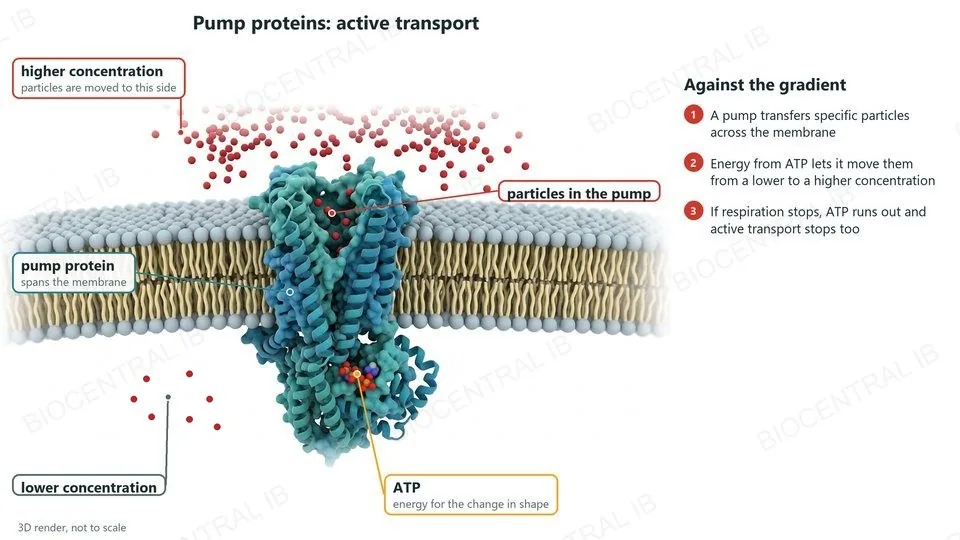
Pump proteins carry out active transport.
  • Pumps use energy from ATP to transfer specific particles across the membrane.
  • Because energy is used, pumps can move particles against a concentration gradient, from a lower to a higher concentration.
  • If respiration stops, the supply of ATP stops, and so does active transport.
CompareChannels: down the gradient, no ATP. Pumps: can go against it, using ATP.
A 3D render of a pump protein spanning a membrane, with ATP bound on the cytoplasm side and particles inside the pump. Energy from ATP moves the particles from the lower to the higher concentration, where many have collected
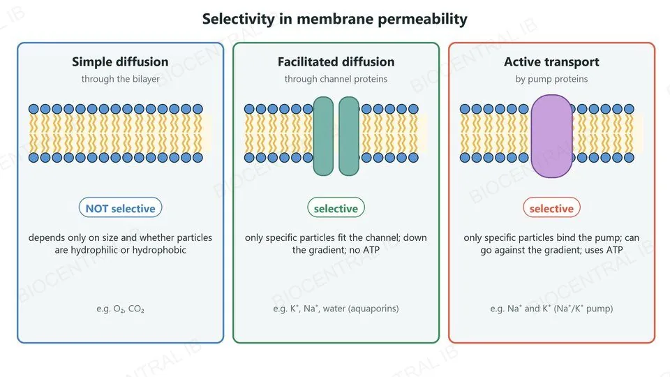
Three panels: simple diffusion through the bilayer is not selective; facilitated diffusion through channels and active transport by pumps are selective
B2.1.8

Selective permeability

Facilitated diffusion and active transport make membranes selectively permeable.
  • Channels and pumps are specific: each lets through only particular ions or molecules.
  • Permeability by simple diffusion is not selective.
  • Whether a particle crosses by simple diffusion depends only on its size and on whether it is hydrophilic or hydrophobic.
Why it mattersBy choosing which proteins to put in a membrane, a cell controls what crosses it.
Part three

The membrane as a whole

B2.1.9 – B2.1.10
B2.1.9

Glycoproteins and glycolipids

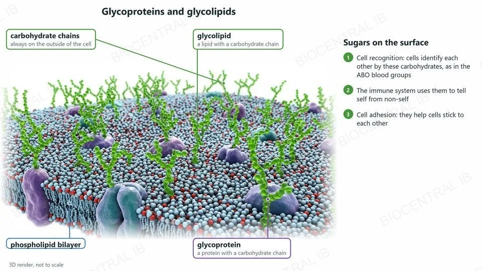
Glycoproteins and glycolipids are proteins and lipids in the membrane with carbohydrate structures linked to them.
  • The carbohydrate parts are always on the extracellular side of the membrane.
  • Cell recognition: cells identify each other by these carbohydrates; for example, the immune system distinguishes self from non-self, and the ABO blood groups depend on them.
  • Cell adhesion: they help cells stick to each other.
LinkThe ABO antigens are in B1.1.
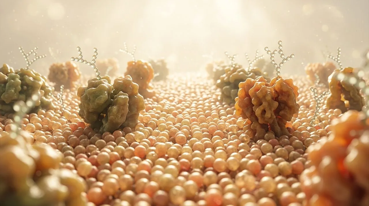
A 3D render of the outer surface of a membrane: green carbohydrate chains stick out on the outside of the cell, attached to proteins (glycoproteins) and to lipids (glycolipids). They allow cell recognition, as in the ABO blood groups, and cell adhesion
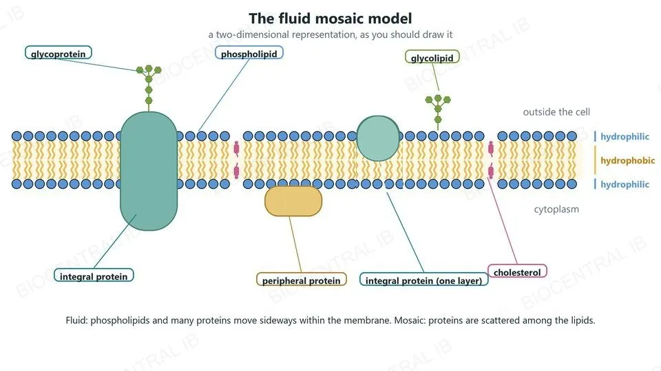
A labelled two-dimensional fluid mosaic model: phospholipid bilayer, integral proteins, a peripheral protein, a glycoprotein, a glycolipid and cholesterol, with the hydrophilic surfaces and hydrophobic core marked
B2.1.10

The fluid mosaic model

The fluid mosaic model describes the structure of membranes.
  • Fluid: phospholipids and many proteins can move sideways within the membrane.
  • Mosaic: proteins are scattered among the lipids, like tiles in a mosaic.
  • Draw it in two dimensions with phospholipids, integral and peripheral proteins, glycoproteins and cholesterol, and mark the hydrophilic and hydrophobic regions.
SkillPractise drawing this diagram: it is often asked for in exams.
Quick check

A membrane made only of phospholipids, with no proteins, separates two solutions. Which substance crosses it most easily?

sodium ions
oxygen
glucose
amino acids
Correct answer: oxygen. It is small and non-polar, so it passes between the phospholipids by simple diffusion. Ions are charged and glucose and amino acids are polar, so they need transport proteins.
Part four · HL

Fluidity and specialized transport

B2.1.11 – B2.1.17 · HL only
B2.1.11 · HL

Fatty acids and fluidity

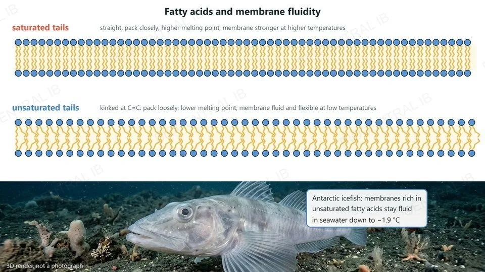
The fatty acids in a bilayer affect how fluid it is.
  • Unsaturated fatty acids have kinks at their C=C bonds, so they pack loosely and have lower melting points: membranes stay fluid and flexible at the temperatures a cell experiences.
  • Saturated fatty acids are straight, pack closely and have higher melting points, so they make membranes stronger at higher temperatures.
  • Antarctic icefish live in seawater at about −1.9 °C; their membranes are rich in unsaturated fatty acids.
AdaptationMembrane composition matches the temperature of the habitat.
Two bilayers: straight saturated tails packed closely, and kinked unsaturated tails packed loosely; below, an Antarctic icefish whose membranes are rich in unsaturated fatty acids
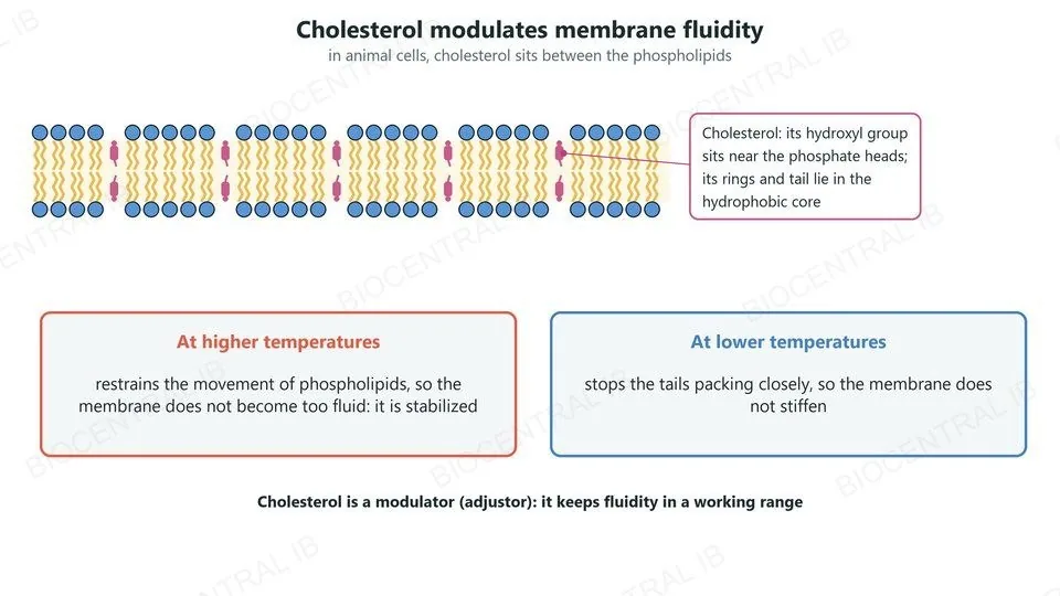
A bilayer with cholesterol molecules between the phospholipids; at higher temperatures cholesterol restrains phospholipid movement, and at lower temperatures it stops the tails packing closely
B2.1.12 · HL

Cholesterol and fluidity

In animal cells, cholesterol molecules sit between the phospholipids of the membrane.
  • Its small hydroxyl group lies near the phosphate heads; its rings and tail lie in the hydrophobic core.
  • At higher temperatures cholesterol restrains the movement of phospholipids, stabilizing the membrane.
  • At lower temperatures it stops the tails packing closely, so the membrane does not stiffen.
RememberCholesterol is a modulator: it adjusts fluidity both ways.
B2.1.13 · HL

Endocytosis

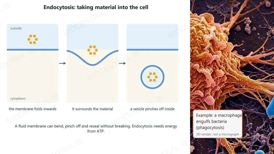
Membrane fluidity allows vesicles to form and to fuse with membranes.
  • In endocytosis, part of the plasma membrane folds inwards around material outside the cell.
  • The membrane pinches off to form a vesicle inside the cell, and the bilayer reseals without breaking.
  • Example: a macrophage engulfs bacteria (phagocytosis); cells also take in cholesterol carried in LDL particles this way.
RememberEndocytosis uses energy from ATP.
Three stages of endocytosis: the membrane folds inwards around material, then a vesicle pinches off inside the cell; beside it a macrophage engulfing bacteria
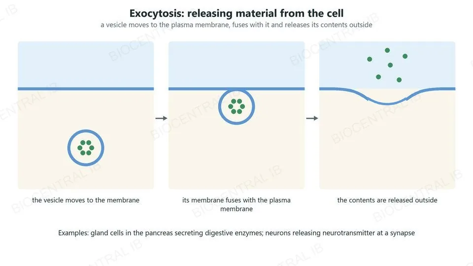
Three stages of exocytosis: a vesicle moves to the plasma membrane, fuses with it and releases its contents outside the cell
B2.1.13 · HL

Exocytosis

In exocytosis, material leaves the cell in vesicles.
  • A vesicle moves to the plasma membrane, and the two membranes fuse.
  • The contents of the vesicle are released outside the cell, and its membrane becomes part of the plasma membrane.
  • Examples: gland cells in the pancreas secrete digestive enzymes; neurons release neurotransmitter at synapses.
LinkProteins for secretion are made on the rough ER and packaged by the Golgi apparatus (B2.2).
B2.1.14 · HL

Gated ion channels in neurons

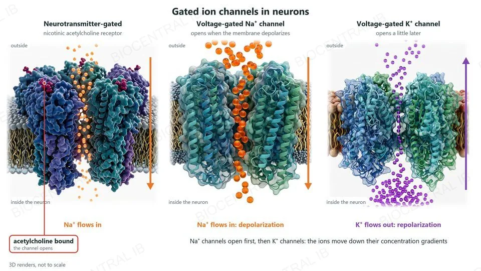
Gated ion channels open only in response to a particular signal.
  • Neurotransmitter-gated: the nicotinic acetylcholine receptor opens when acetylcholine binds to it, and Na⁺ flows into the neuron.
  • Voltage-gated: sodium and potassium channels open when the membrane potential changes.
  • Na⁺ channels open first, so Na⁺ flows in (depolarization); then K⁺ channels open, so K⁺ flows out (repolarization).
LinkNerve impulses and synapses are in C2.2.
Three 3D renders of gated channels in a neuron membrane. A neurotransmitter-gated channel, the nicotinic acetylcholine receptor, opens when acetylcholine binds and Na⁺ flows in. A voltage-gated Na⁺ channel lets Na⁺ flow in: depolarization. A voltage-gated K⁺ channel, opening a little later, lets K⁺ flow out: repolarization
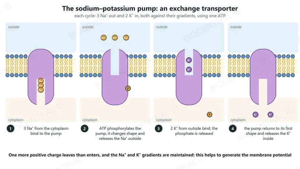
Four steps of the sodium–potassium pump: 3 Na⁺ bind from the cytoplasm; ATP phosphorylates the pump, which changes shape and releases Na⁺ outside; 2 K⁺ bind from outside and the phosphate is released; the pump returns to its first shape and releases K⁺ inside
B2.1.15 · HL

The sodium–potassium pump

Sodium–potassium pumps are exchange transporters: they move two ions in opposite directions.
  • In each cycle, the pump uses one ATP to move 3 Na⁺ out of the cell and 2 K⁺ into it, both against their concentration gradients.
  • ATP phosphorylates the pump, changing its shape; releasing the phosphate changes it back.
  • The pumps keep Na⁺ high outside and K⁺ high inside, and move more positive charge out than in, generating membrane potentials.
Why it mattersNeurons depend on these gradients for their resting potential.
B2.1.16 · HL

Sodium-dependent glucose cotransport

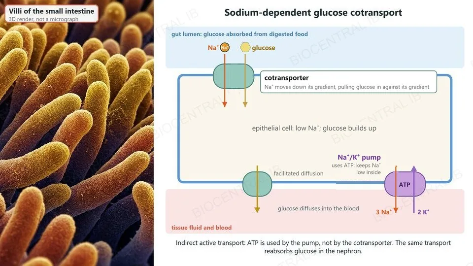
Sodium-dependent glucose cotransporters are an example of indirect active transport.
  • Na⁺/K⁺ pumps use ATP to keep the Na⁺ concentration inside the cell low.
  • The cotransporter lets Na⁺ diffuse into the cell, down its gradient, and carries glucose in with it, against the glucose gradient.
  • These cotransporters absorb glucose into cells lining the small intestine and reabsorb glucose into cells of the nephron.
RememberIndirect: the energy comes from the Na⁺ gradient, which the pump made using ATP.
Villi of the small intestine beside an epithelial cell: a cotransporter on the gut side moves Na⁺ down its gradient and glucose with it; a Na⁺/K⁺ pump on the blood side keeps Na⁺ low inside; glucose diffuses into the blood
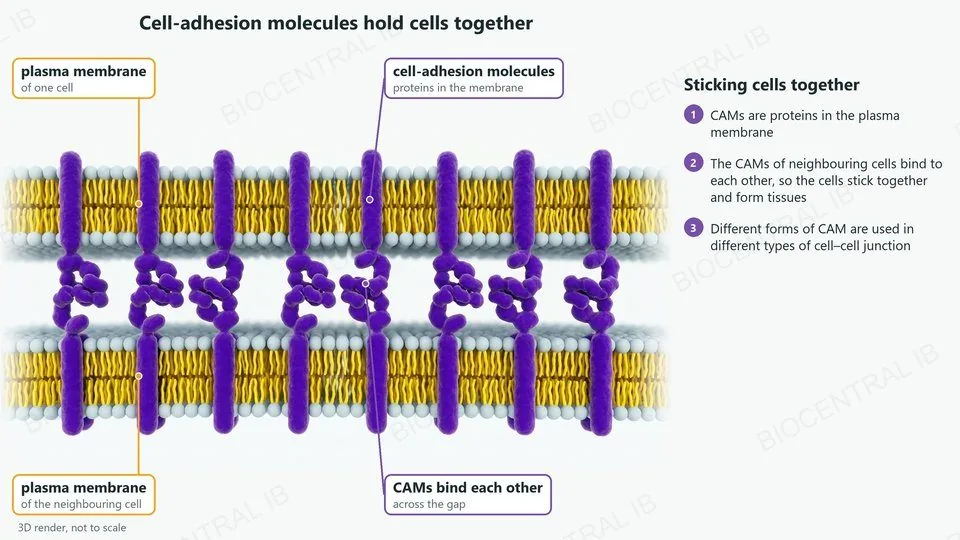
A 3D render of the plasma membranes of two neighbouring cells. Cell-adhesion molecules, proteins in each membrane, reach across the gap and bind to each other, holding the cells together
B2.1.17 · HL

Cells adhere to form tissues

Cell-adhesion molecules (CAMs) hold cells together to form tissues.
  • CAMs are proteins in the plasma membrane.
  • The CAMs of neighbouring cells bind to each other, sticking the cells together.
  • Different forms of CAM are used for different types of cell–cell junction; you do not need to know the details of the different CAMs or junctions.
Why it mattersWithout adhesion, cells could not build tissues and organs.
Quick check · HL

A drug stops the sodium–potassium pumps of intestinal cells. Why does glucose absorption by the sodium-dependent cotransporters soon stop as well?

the drug blocks the glucose cotransporter itself
Na⁺ builds up inside, so the Na⁺ gradient is lost
glucose now diffuses out through the pump
the cotransporter uses ATP from the pump
Correct answer: the Na⁺ gradient is lost. Without the pumps, Na⁺ that enters with glucose is not removed, so the Na⁺ concentration inside rises. With no gradient for Na⁺ to move down, the cotransporter can no longer carry glucose in.

Key vocabulary

Worth being able to define in a single sentence each

Amphipathic
having a hydrophilic part and a hydrophobic part.
Phospholipid bilayer
two layers of phospholipids, tails inwards.
Simple diffusion
net movement down a gradient, directly through the bilayer.
Integral protein
a protein embedded in one or both lipid layers.
Peripheral protein
a protein attached to one surface of the membrane.
Osmosis
net movement of water to a higher solute concentration.
Aquaporin
a channel protein that lets water cross rapidly.
Facilitated diffusion
passive movement through a channel protein.
Active transport
movement by a pump, using ATP, against a gradient.
Glycoprotein
a membrane protein with carbohydrate attached.

Key vocabulary · HL

Additional higher level terms

Membrane fluidity
how freely the lipids and proteins of a membrane move.
Cholesterol
a lipid that modulates the fluidity of animal membranes.
Endocytosis
taking material in by forming a vesicle from the membrane.
Exocytosis
releasing material when a vesicle fuses with the membrane.
Gated channel
a channel that opens only in response to a signal.
Exchange transporter
a pump that moves two ions in opposite directions.
Cotransporter
a protein that moves two substances together, one down its gradient.
CAM
cell-adhesion molecule: a membrane protein that binds cells together.

Where this shows up again

B1.1 · Carbohydrates and lipids
How does the amphipathic structure of phospholipids cause them to form bilayers in water?
B2.2 · Organelles and compartmentalization
How do vesicles from the Golgi apparatus deliver proteins to the plasma membrane by exocytosis?
C2.2 · Neural signalling
How do sodium–potassium pumps and voltage-gated channels together produce a nerve impulse?
D2.3 · Water potential
How is osmosis described using water potential?

B2.1 Membranes and membrane transport — one-page recap

Screenshot this slide to revise from

Bilayer
  • Amphipathic phospholipids form bilayers in water.
  • Hydrophobic core: a barrier to ions, polar and large molecules.
Passive transport
  • Simple diffusion: O₂, CO₂ between phospholipids.
  • Osmosis (aquaporins speed it up); channels for facilitated diffusion.
Proteins and selectivity
  • Integral (embedded) and peripheral (on the surface).
  • Channels and pumps are selective; simple diffusion is not.
Surface and model
  • Glycoproteins and glycolipids: carbohydrate outside; recognition and adhesion.
  • Fluid mosaic model: draw it with all five components.
HL · Fluidity and vesicles
  • Unsaturated: fluid when cold; saturated: strong when hot; cholesterol modulates.
  • Fluidity allows endocytosis and exocytosis.
HL · Specialized transport
  • Gated channels (ACh receptor; voltage-gated Na⁺, K⁺); Na⁺/K⁺ pump: 3 out, 2 in.
  • Na⁺-glucose cotransport (gut, nephron); CAMs make tissues.
Next step

Now test yourself on B2.1

Reading the slides is half the job. The other half is answering questions the way the exam asks them.

52 quiz questions8 data questions5 exam questionsmarkschemes included

Unlock every lesson with Pro — quizzes, data questions and exam papers. Two lessons are completely free to try: A1.1 Water and B1.1 Carbohydrates and lipids.

One-page summary, key terms and sample questions for B2.1 →

A membrane is a barrier with doors.

The lipids keep things out; the proteins decide what gets through.
B2.1 Membranes and membrane transport · BioCentral IB
Use ↓ ↑ or click to navigate
01 / 20