BioCentral IBBioCentral IB
IB Biology · Theme A · A2.2

Cell Structure

The unit of life — and how we see inside it
Guiding questions

What are the features common to all cells and the features that differ?

How is microscopy used to investigate cell structure?

Part one

The cell as the unit of life

A2.2.1 · A2.2.4
A2.2.1

Every organism is built from cells

All living organisms are composed of one or more cells, and cells are the basic units of structure.
  • From a single bacterium to the roughly 30 trillion cells of a human body, every organism is made of cells.
  • Tissues, organs and whole organisms are built from cells and the materials cells make, such as wood and bone.
Nature of scienceDeductive reasoning: because cell theory says all organisms are made of cells, we can predict that a newly discovered organism will consist of one or more cells.
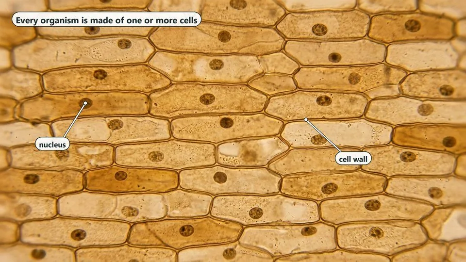
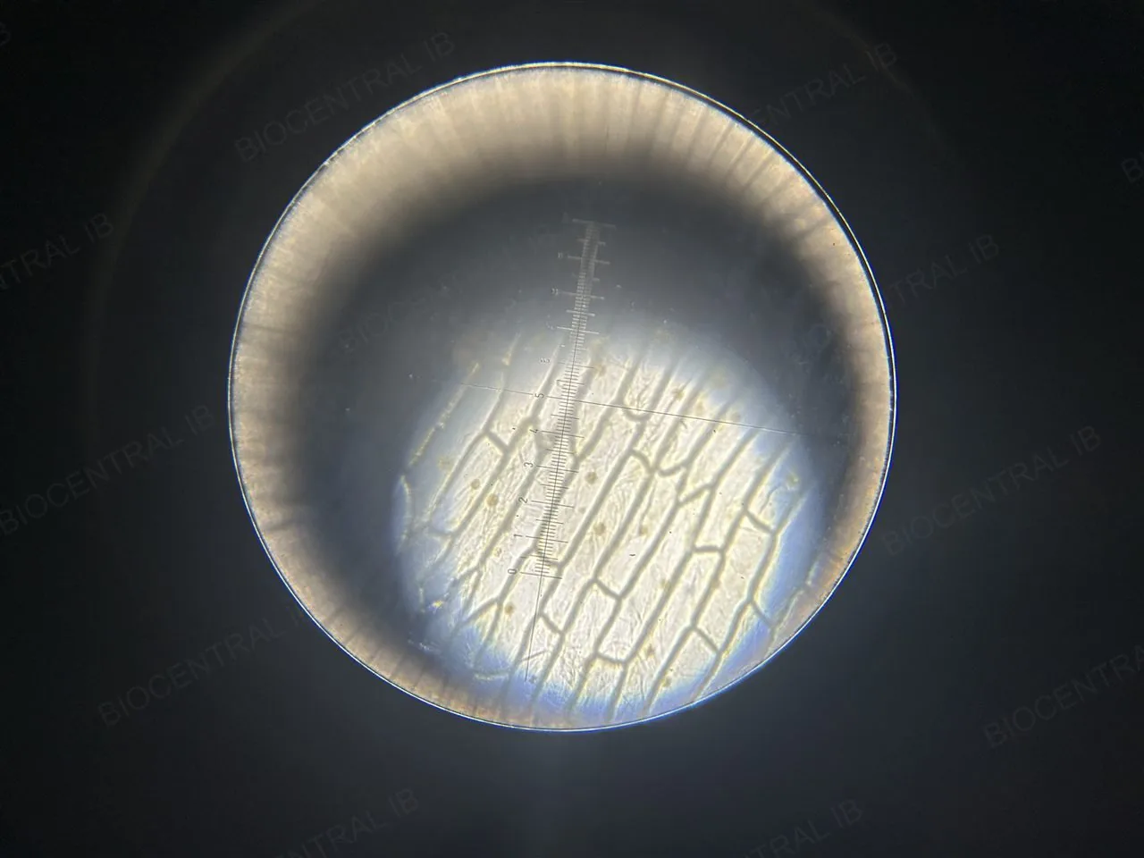
A light micrograph of onion epidermis stained with iodine: a sheet of long, brick-shaped cells fitted together, each with a brown cell wall and one darker nucleus. Labels point to a cell wall and a nucleus
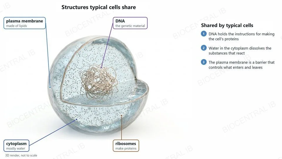
A 3D render of a simple round cell cut open: a thin plasma membrane made of lipids, a watery cytoplasm, a tangled loop of DNA, the genetic material, and many tiny ribosomes that make proteins. The membrane is a barrier that controls what enters and leaves, and water in the cytoplasm dissolves the substances that react
A2.2.4

Structures typical cells share

Typical cells have DNA as their genetic material, and a cytoplasm made mainly of water enclosed by a plasma membrane made of lipids.
  • DNA holds the instructions for making the cell’s proteins, and is passed on when the cell divides.
  • Water in the cytoplasm dissolves the substances the cell needs, so its chemical reactions can happen.
  • The lipids of the plasma membrane are hydrophobic, so it is a barrier between the cell and its surroundings that controls what enters and leaves.
Also in nearly every cellRibosomes, which make proteins. Everything else, such as a nucleus or a cell wall, is found only in some cells.
Part two

Seeing the invisible: microscopy

A2.2.2 · A2.2.3
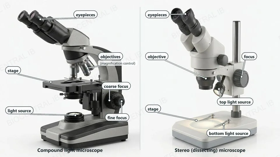
Photographs of the two microscopes in the data booklet with labels. Compound light microscope: eyepieces, objectives (magnification control), stage, coarse focus, fine focus and light source. Stereo (dissecting) microscope: eyepieces, objective, focus, top light source, stage and bottom light source
A2.2.2 · Practical skill

Using a light microscope

  • Make a temporary mount: a thin layer of cells in a drop of water or stain on a slide, under a cover slip.
  • Stains such as iodine or methylene blue make colourless structures, such as nuclei, visible.
  • Focus with the coarse focus at low power, then the fine focus. At higher power, use only the fine focus.
  • Take photographs through the eyepiece or with a camera fitted to the microscope.
Data bookletThe data booklet shows the parts of a compound light microscope and a stereo (dissecting) microscope.
A2.2.2 · Practical skill

Calculating magnification

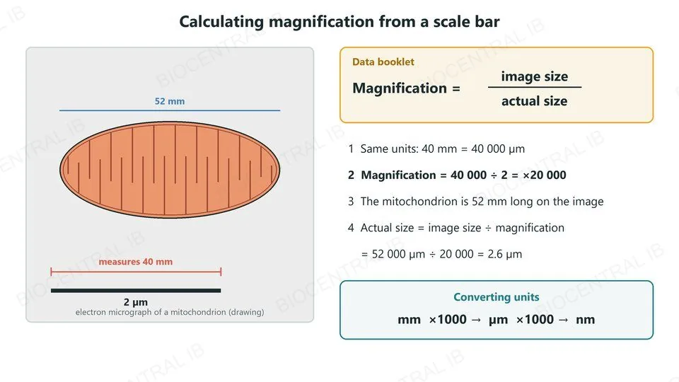
Magnification is how many times larger the image is than the real object: magnification = image size ÷ actual size.
  • A scale bar labelled 2 µm measures 40 mm on a micrograph. 40 mm = 40 000 µm, so magnification = 40 000 ÷ 2 = ×20 000.
  • Rearranged: actual size = image size ÷ magnification. A mitochondrion 52 mm long on that image is 52 000 µm ÷ 20 000 = 2.6 µm long.
Why it mattersThe most common error is mixing units. Convert first: 1 mm = 1000 µm and 1 µm = 1000 nm.
A drawing of a mitochondrion from an electron micrograph, 52 mm long, with a scale bar labelled 2 µm that measures 40 mm. Beside it, the data-booklet formula: magnification equals image size divided by actual size. Steps: 40 mm equals 40 000 µm; magnification equals 40 000 divided by 2, which is times 20 000; the mitochondrion is 52 mm long on the image; actual size equals 52 000 µm divided by 20 000, which is 2.6 µm. A box shows converting units: millimetres times 1000 gives micrometres, times 1000 gives nanometres
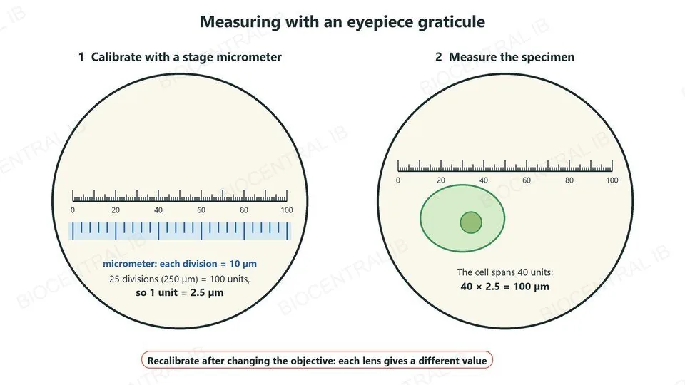
Two circular fields of view. Left, calibration: an eyepiece graticule scale of 100 units lies over a stage micrometer whose divisions are each 10 µm; 25 divisions, 250 µm, equal 100 units, so 1 unit is 2.5 µm. Right, measurement: a cell spans 40 graticule units, so it is 40 times 2.5, which is 100 µm long. A note says to recalibrate after changing the objective
A2.2.2 · Practical skill

Measuring with a graticule

  • An eyepiece graticule is a scale inside the eyepiece. Its units mean nothing until it is calibrated.
  • Calibrate it against a stage micrometer, a slide with a scale of known length, separately for each objective.
  • Measure the specimen in graticule units, then convert to µm.
  • A scale bar on a drawing or photograph shows the real length a line represents, so it stays correct if the image is resized.
Nature of scienceMeasuring with an instrument is quantitative observation: it turns what we see into numbers that can be compared and checked.
A2.2.3

Resolution and electron microscopes

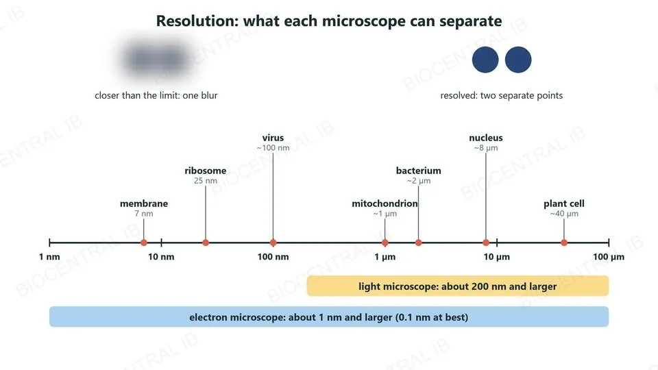
Resolution is the smallest distance between two points at which they can still be seen as separate.
  • A light microscope resolves to about 200 nm. Electron microscopes use beams of electrons, whose wavelength is far shorter than light’s, so they resolve to about 1 nm or better.
  • Electron microscopes reveal ultrastructure such as membranes, ribosomes and cristae, but specimens must be dead, fixed and in a vacuum, and images are black and white.
Why it mattersMagnifying beyond the resolution limit only makes a bigger blur.
Top, two dots so close that they blur into one, and two dots resolved as separate points. Below, a scale from 1 nm to 100 µm marking a membrane 7 nm, a ribosome 25 nm, a virus about 100 nm, a mitochondrion about 1 µm, a bacterium about 2 µm, a nucleus about 8 µm and a plant cell about 40 µm. A yellow bar shows the light microscope resolving about 200 nm and larger; a blue bar shows the electron microscope resolving about 1 nm and larger, 0.1 nm at best
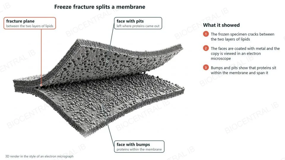
A 3D render, in the style of an electron micrograph, of a membrane split along the fracture plane between its two layers of lipids. The lower face is covered in bumps, proteins within the membrane; the upper face shows the pits they left. The faces are coated with metal and the copy is viewed in an electron microscope, showing that proteins sit within the membrane and span it
A2.2.3

Freeze fracture: splitting the membrane

Freeze fracture freezes a specimen and then cracks it, so membranes split along the weak plane between their two layers of lipids.
  • The exposed faces are coated with a thin layer of metal, and this copy is viewed with an electron microscope.
  • One face shows bumps (proteins); the other shows the pits they left.
Main advantageIt shows the inside of a membrane: proteins span the lipid bilayer rather than coating its surfaces, which supports the fluid mosaic model.
A2.2.3

Cryo-EM: freezing molecules mid-shape

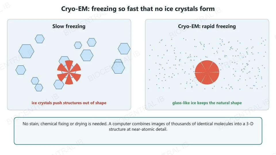
Cryogenic electron microscopy (cryo-EM) freezes a specimen so fast that no ice crystals form.
  • Ordinary freezing makes ice crystals that distort delicate structures; in cryo-EM the water becomes glass-like ice instead.
  • No stain, chemical fixing or drying is needed.
  • A computer combines images of thousands of identical molecules into a 3-D structure at near-atomic detail.
Main advantageProteins and viruses are seen close to their natural, hydrated shape.
Two panels. Slow freezing: hexagonal ice crystals form and push a round molecule out of shape. Cryo-EM, rapid freezing: glass-like ice with no crystals, and the molecule keeps its natural shape. A note says no stain, fixing or drying is needed, and a computer combines images of thousands of identical molecules into a 3-D structure
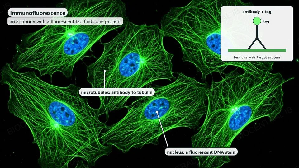
A fluorescence micrograph of several flat animal cells: a fine network of microtubules glows green in each cell, labelled antibody to tubulin, and each nucleus glows blue, labelled a fluorescent DNA stain. An inset shows a Y-shaped antibody with a glowing tag binding only its target protein
A2.2.3

Fluorescent stains and immunofluorescence

  • A fluorescent stain binds one kind of molecule, such as DNA, and glows when lit with light of the right wavelength.
  • In immunofluorescence, a fluorescent tag is attached to an antibody that binds only one target protein, so the glow shows exactly where that protein is.
  • Using different colours, several molecules can be shown in the same cell.
Main advantageSpecific molecules can be located inside cells with a light microscope.
Part three

Prokaryotes, eukaryotes, and single-celled life

A2.2.5 · A2.2.6 · A2.2.7
A2.2.5

Prokaryote cell structure

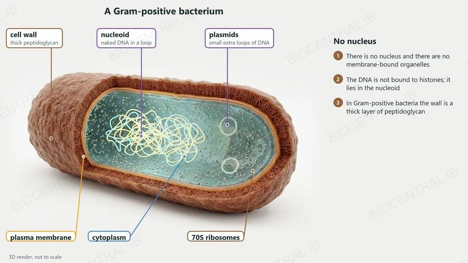
A prokaryotic cell has no nucleus and no membrane-bound organelles.
  • Its genetic material is naked DNA in a loop, not bound to histones, in a region called the nucleoid. Its ribosomes are 70S.
  • A cell wall outside the plasma membrane gives shape and protection. In Gram-positive bacteria such as Bacillus and Staphylococcus it is a thick layer of peptidoglycan.
  • Many bacteria also have plasmids, small extra loops of DNA.
Why it mattersProkaryote structure varies between species; you need the Gram-positive pattern, not every variation.
A 3D render of a rod-shaped Gram-positive bacterium cut open: a thick cell wall of peptidoglycan, the plasma membrane inside it, cytoplasm, the nucleoid, a tangled loop of naked DNA, two small plasmids and many 70S ribosomes. There is no nucleus and there are no membrane-bound organelles
A2.2.6

Eukaryote cell structure

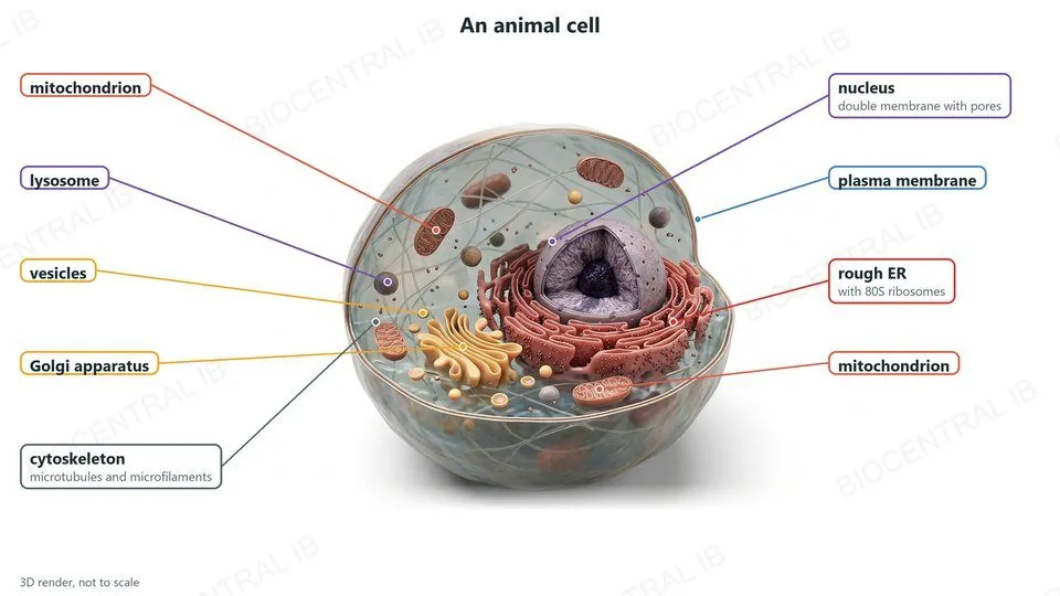
A eukaryotic cell (animals, plants, fungi, protists) has a nucleus and membrane-bound organelles.
  • Its DNA is linear, associated with histone proteins, and enclosed within the nucleus's own double membrane, which has pores.
  • Membrane-bound organelles — the mitochondrion, endoplasmic reticulum, Golgi apparatus and vesicles (including lysosomes) — each provide their own internal environment suited to a specific job, an arrangement called compartmentalisation.
  • Its ribosomes are 80S, larger than a prokaryote's 70S.
  • A cytoskeleton of microtubules and microfilaments supports the cell and its contents.
Why it mattersCompartmentalisation lets incompatible reactions run simultaneously in the same cell, each isolated in its own organelle.
A 3D render of an animal cell cut open. Labels: nucleus, with a double membrane with pores; rough ER with 80S ribosomes; Golgi apparatus; vesicles; a lysosome; two mitochondria; the cytoskeleton of microtubules and microfilaments; and the plasma membrane
A2.2.7

One cell doing everything

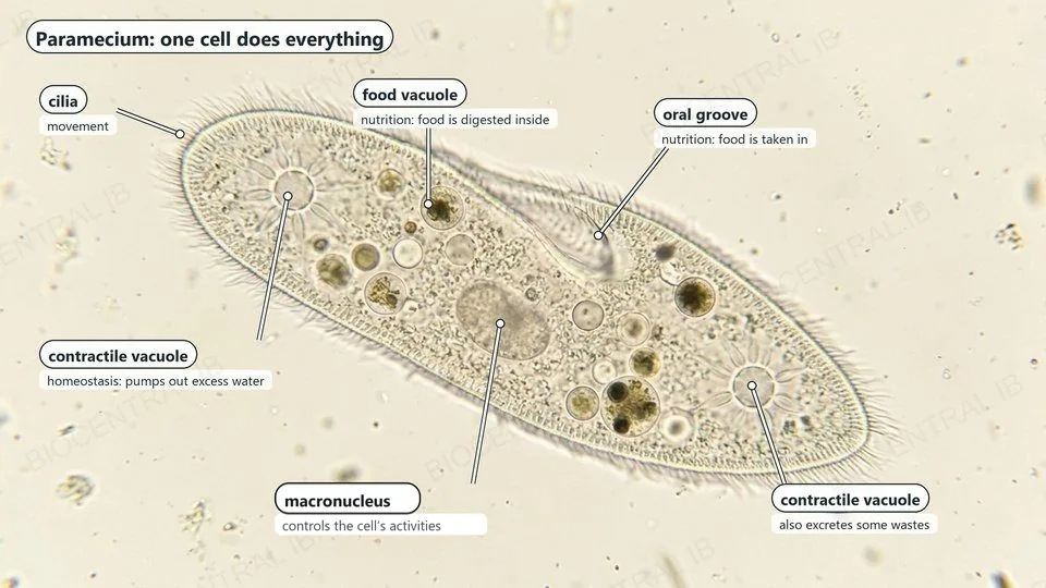
A unicellular organism, such as Paramecium, Amoeba or Chlamydomonas, carries out every process of life in one cell.
  • In Paramecium, cilia give movement; an oral groove and food vacuoles give nutrition; contractile vacuoles pump out excess water for homeostasis and excretion.
  • Its enzymes carry out metabolism; it grows, reproduces by dividing, and responds to stimuli by changing direction.
Why it mattersThere are no other cells to share the work, so every process happens inside one cell.
A light micrograph of a slipper-shaped Paramecium with labels: cilia for movement; contractile vacuoles that pump out excess water (homeostasis) and also excrete some wastes; food vacuoles where food is digested; the oral groove where food is taken in; the macronucleus, which controls the cell's activities
Quick check

A student measures a scale bar as 20 mm long on a micrograph. The scale bar is labelled 2 μm. What is the magnification?

×10
×10,000
×40
×2,000
Correct answer: ×10,000. Convert to the same unit first (20 mm = 20,000 μm), then divide: 20,000 ÷ 2 = 10,000. Skipping the unit conversion — dividing 20 by 2 — is the most common version of this mistake.
Part four

Diversity, exceptions, and reading cells

A2.2.8 · A2.2.9 · A2.2.10 · A2.2.11
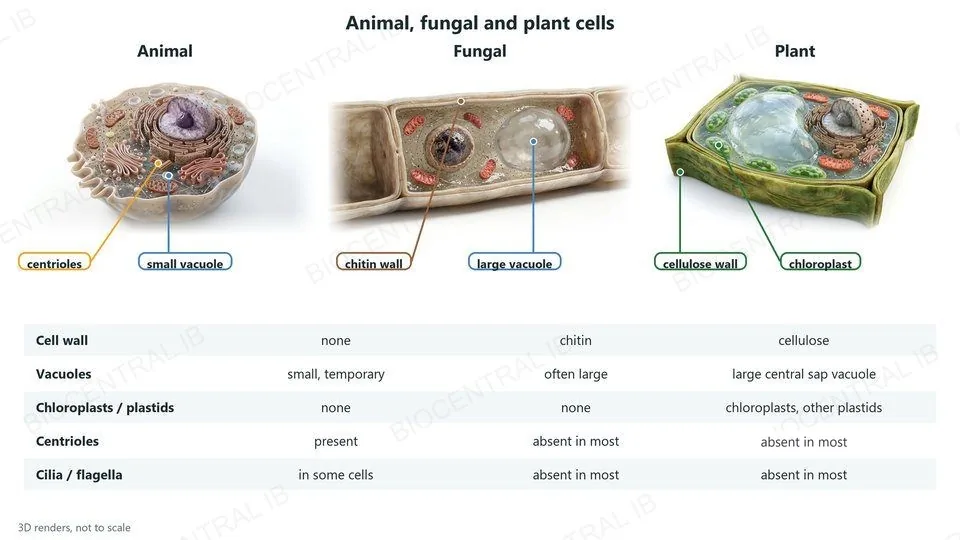
Three 3D renders: an animal cell with centrioles and a small vacuole; a fungal cell in a hypha with a chitin wall and a large vacuole; and a plant cell with a cellulose wall, a large central vacuole and chloroplasts. A table compares them. Cell wall: none, chitin, cellulose. Vacuoles: small and temporary, often large, a large central sap vacuole. Chloroplasts and plastids: none, none, chloroplasts and other plastids. Centrioles: present, absent in most, absent in most. Cilia and flagella: in some cells, absent in most, absent in most
A2.2.8

Animal, fungal and plant cells

  • Cell walls: none in animal cells, chitin in fungi, cellulose in plants.
  • Vacuoles: small and temporary in animal cells; a large permanent sap vacuole in plant cells, which stores water and keeps the cell firm. Fungal cells often have large vacuoles.
  • Plastids: only plant cells have chloroplasts and other plastids.
  • Centrioles, cilia and flagella occur in animal cells but are absent from most plant and fungal cells.
Why it mattersThese differences sit on top of the shared eukaryotic plan from A2.2.6.
A2.2.9

Red blood cells lose their nucleus

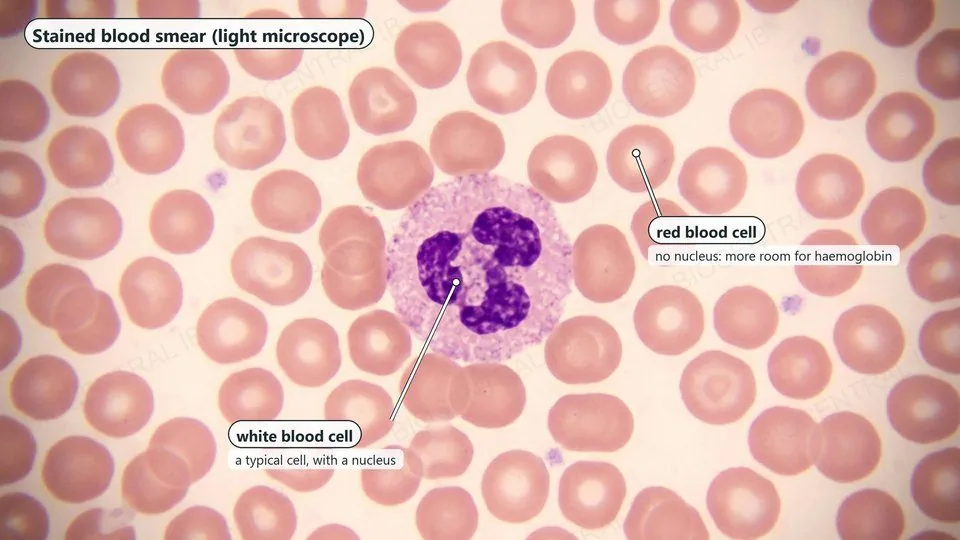
A mature mammalian red blood cell has no nucleus — one clear example of atypical cell structure defined by the number of nuclei a cell contains.
  • As a red blood cell matures in the bone marrow, it expels its nucleus along with most other organelles, becoming a flattened, biconcave disc almost entirely filled with haemoglobin.
  • The extra internal space raises the amount of haemoglobin — and so the amount of oxygen — each cell can carry, while the biconcave shape increases surface area for gas exchange.
Why it mattersLosing its nucleus means a mature red blood cell can no longer divide or make new proteins — it survives roughly 120 days before the body replaces it.
A stained blood smear under a light microscope: many round pale pink red blood cells with no nucleus, which leaves more room for haemoglobin, and one white blood cell with a purple lobed nucleus
A2.2.9 continued

Sieve tubes lose their nucleus but stay alive

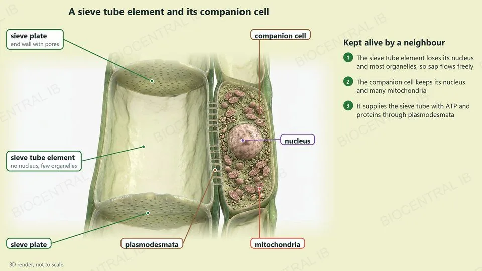
A phloem sieve-tube element also loses its nucleus at maturity — but unlike a red blood cell, it survives only because a neighbouring cell keeps it alive.
  • Losing its nucleus and most organelles clears the tube's interior, letting sugars (mainly sucrose) flow through pores in its end walls, called sieve plates, with minimal obstruction.
  • An adjacent, fully nucleate companion cell stays connected to the sieve-tube element through plasmodesmata, supplying it with ATP and proteins it can no longer make for itself.
Why it mattersA sieve-tube element cannot survive alone — its atypical structure only works because of this permanent partnership with its companion cell.
A 3D render of a sieve tube element beside its companion cell, cut open. The sieve tube element is a wide tube with a sieve plate, an end wall with pores, at each end, and has no nucleus and few organelles, so sap flows freely. The narrower companion cell has a nucleus and many mitochondria, and is joined to the sieve tube by plasmodesmata, through which it supplies ATP and proteins
A2.2.9 continued

One giant cell, many nuclei

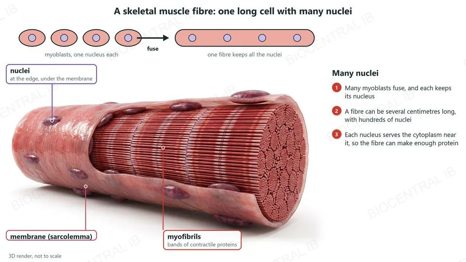
A skeletal muscle fibre is a single, unusually large cell containing many nuclei, formed by the fusion of many separate embryonic cells into one.
  • During development, many individual myoblasts fuse together into one continuous fibre — and each keeps its own nucleus rather than the fibre reducing to just one.
  • A fibre can run centimetres long and carry hundreds of nuclei spread along its length, each supporting protein synthesis in the section of cytoplasm nearest to it.
Why it mattersA muscle fibre needs far more protein-making capacity than a single nucleus could support — multiple nuclei let it sustain its huge volume of contractile protein.
Top, four myoblasts with one nucleus each fuse into one fibre that keeps all the nuclei. Below, a 3D render of a skeletal muscle fibre cut open: many nuclei at the edge, under the membrane (sarcolemma), and the inside packed with myofibrils, bands of contractile proteins. A fibre can be several centimetres long with hundreds of nuclei
A2.2.9 continued

No walls between nuclei

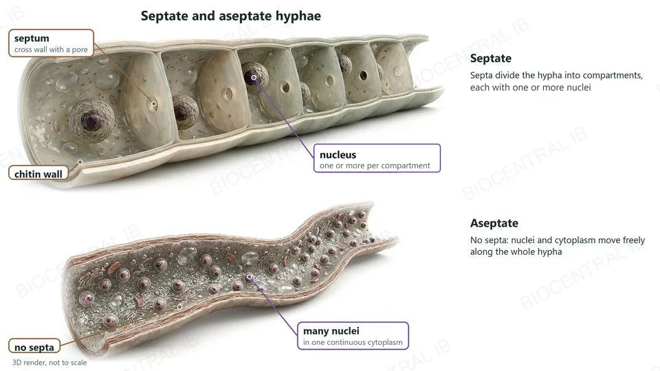
Aseptate fungal hyphae are long tubes of continuous cytoplasm containing many nuclei, with no cross-walls dividing one region from the next.
  • Most fungal hyphae are divided into compartments by cross-walls called septa; aseptate hyphae lack these, so nuclei and cytoplasm move freely along the whole length of the hypha.
  • This uninterrupted structure lets resources and signalling molecules travel rapidly through the fungus, supporting fast growth at the hyphal tip.
Why it mattersAseptate hyphae, like the red blood cell, phloem sieve-tube element and muscle fibre, show that nucleus number is set by function, not by a fixed one-nucleus-per-cell rule.
Two 3D renders of fungal hyphae cut lengthways. Top, a septate hypha: cross walls called septa, each with a pore, divide the tube into compartments with one or more nuclei each, inside a chitin wall. Bottom, an aseptate hypha: no septa, so many nuclei share one continuous cytoplasm and move freely along the hypha
A2.2.10

Reading an electron micrograph

Identify structures by their shape, their membranes and their size.
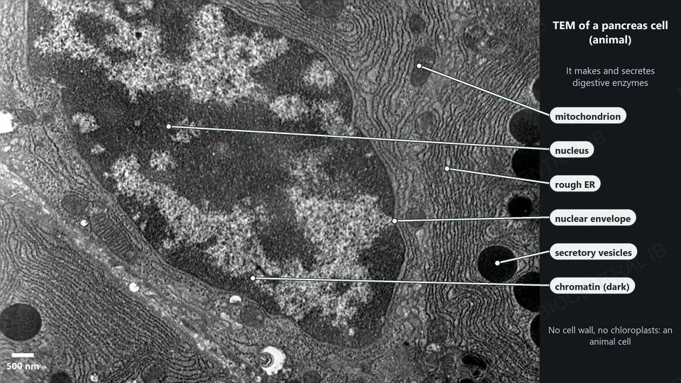
  • Nucleus: large, with a double membrane with pores and dark patches of chromatin.
  • Rough ER: parallel membranes studded with ribosomes.
  • Mitochondrion: a double membrane, the inner one folded into cristae.
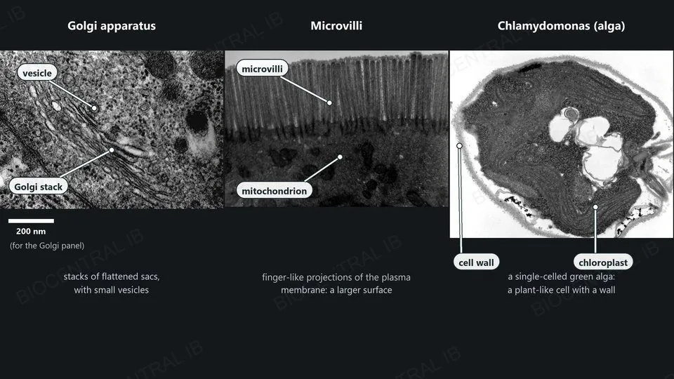
  • Golgi apparatus: a stack of curved, flattened sacs with vesicles.
Scale is a clueA structure a few nanometres across cannot be a nucleus; one several micrometres across cannot be a ribosome.
A transmission electron micrograph of a pancreas cell with a 500 nm scale bar. Labels point to the nucleus with dark patches of chromatin, the nuclear envelope, parallel membranes of rough ER, a mitochondrion and dark round secretory vesicles. A note says there is no cell wall and no chloroplasts, so this is an animal cell
Louisa Howard, Dartmouth Electron Microscope Facility · Public domain
Three transmission electron micrographs. The Golgi apparatus: a stack of curved, flattened sacs with small vesicles. Microvilli: finger-like projections of the plasma membrane on the surface of a cell, with mitochondria below. Chlamydomonas, a single-celled green alga: a cell wall around the cell and a chloroplast full of thylakoid membranes
Louisa Howard, Katherine Connolly; Dartmouth EM Facility · Public domain
A2.2.10

Prokaryote, plant or animal?

  • Prokaryote: small (about 1–5 µm), no nucleus, a nucleoid region of DNA, a cell wall and 70S ribosomes.
  • Plant cell: a cell wall, chloroplasts and often a large sap vacuole.
  • Animal cell: no cell wall or chloroplasts; some have microvilli on their surface.
MicrovilliFinger-like projections of the plasma membrane that increase its surface area, as on cells lining the intestine.
A2.2.10

A plant cell in a micrograph

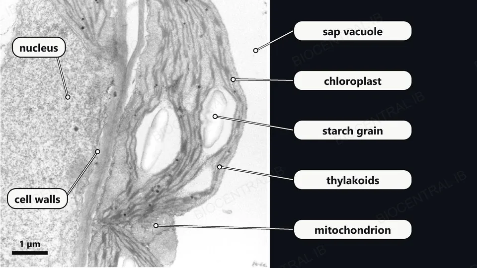
A wall, chloroplasts and a large pale vacuole mark out a plant cell.
  • Cell wall: a thick layer outside the plasma membrane. Two neighbouring cells share a band of two walls.
  • Chloroplast: large, with a double membrane, parallel thylakoids and pale oval starch grains.
  • Sap vacuole: a pale, almost empty region that takes up much of the cell.
  • Mitochondrion: much smaller than a chloroplast, with a double membrane.
Use the scaleThe bar shows 1 µm, so this chloroplast is more than 5 µm long. Measure before you name.
A real transmission electron micrograph of parts of two neighbouring plant cells with a 1 µm scale bar. Labels point to the nucleus, the cell walls of the two cells, a chloroplast with internal thylakoid membranes and two pale starch grains, a mitochondrion and the pale, almost empty sap vacuole
Simon et al. 2013, PLOS ONE 8(6):e65811 · CC BY · lettering replaced
A2.2.11 · Practical skill

Drawing what you actually see

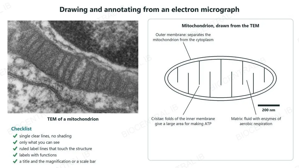
A scientific drawing records only what is visible in one specific micrograph.
  • Draw single, clear, unbroken lines, with no shading, and keep the proportions accurate.
  • Use ruled label lines that touch the structure and never cross.
  • Add a title and the magnification or a scale bar.
AnnotationsEach label needs the structure’s function, e.g. cristae: a large surface area for producing ATP.
Left, an electron micrograph of an elongated mitochondrion. Right, a single-line drawing of it with a scale bar: an outer membrane, an inner membrane folded into cristae, and the matrix, each annotated with its function. A checklist: single clear lines with no shading, only what you can see, ruled label lines that touch the structure, labels with functions, and a title and the magnification or a scale bar

Key vocabulary — cell structure

Worth being able to define in a single sentence each

Magnification
How many times larger an image is than the real, actual object.
Resolution
The smallest distance between two points at which they can still be seen as separate.
Prokaryotic cell
A cell with no nucleus and no membrane-bound organelles; DNA lies free in the nucleoid.
Eukaryotic cell
A cell with a membrane-bound nucleus and membrane-bound organelles.
Nucleoid
The region of a prokaryotic cell where the circular DNA is located; not membrane-bound.
Compartmentalisation
The division of a eukaryotic cell into separate membrane-bound regions, each with its own environment.
Part five · HL only

Origins and specialisation

A2.2.12 · A2.2.13 · A2.2.14
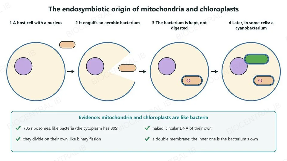
Four steps. 1, a host cell with a nucleus. 2, it engulfs an aerobic bacterium. 3, the bacterium is kept, not digested, inside a second membrane, as a mitochondrion with its own circular DNA. 4, later, in some cells, a cyanobacterium is taken in and becomes a chloroplast. Evidence: 70S ribosomes, naked circular DNA, division on their own like binary fission, and a double membrane
A2.2.12 · HL

Mitochondria were once free-living bacteria

All eukaryotes evolved from a common unicellular ancestor that had a nucleus and reproduced sexually. Mitochondria then evolved by endosymbiosis, and later, in some eukaryotes, chloroplasts did too.
  • An engulfed aerobic bacterium survived inside the host and became the mitochondrion; a cyanobacterium became the chloroplast.
  • Evidence: both have 70S ribosomes, naked circular DNA and the ability to replicate by dividing; both also have a double membrane.
Nature of scienceA theory is strong when it explains many observations and its predictions are supported. Endosymbiosis accounts for a wide range of observations.
A2.2.13 · HL

Same genome, different jobs

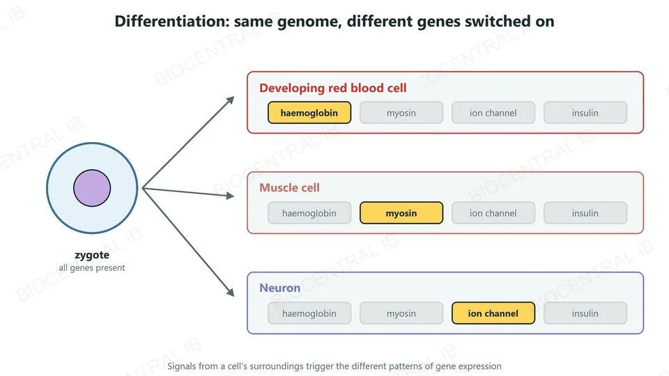
Cell differentiation is the process by which cells become specialised, through different patterns of gene expression.
  • Every cell inherits the same genome from the zygote, but each type expresses a different set of genes: haemoglobin in developing red blood cells, contractile proteins in muscle, ion channels in neurons.
  • Different patterns of gene expression are often triggered by changes in the cell’s environment, such as signals from neighbouring cells.
Why it mattersDifferentiation makes a division of labour between cells possible, so tissues can specialise.
A zygote with all genes present gives rise to three cell types. Each has the same four genes, but only one of these four is switched on: haemoglobin in a developing red blood cell, myosin in a muscle cell and an ion channel gene in a neuron. A note says signals from a cell’s surroundings trigger the different patterns of gene expression
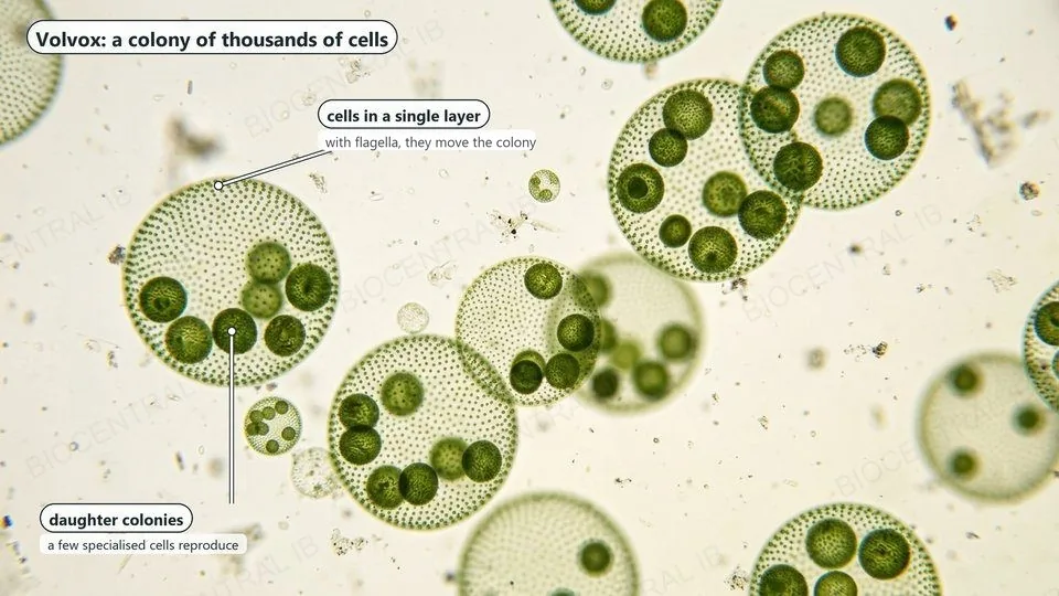
A light micrograph of Volvox colonies: hollow green spheres made of many small cells in a single layer, with darker green daughter colonies inside the larger spheres
A2.2.14 · HL

Multicellularity evolved again and again

Multicellularity has evolved repeatedly: many fungi and eukaryotic algae, and all plants and animals, are multicellular.
  • Cells that stay together after dividing can build a larger body.
  • They can also specialise. In the green alga Volvox, most cells move the colony while a few make new daughter colonies.
AdvantagesA larger body size, and cell specialisation that divides the work between different cells.

Key vocabulary — origins & specialisation

HL only — worth being able to define in a single sentence each

EndosymbiosisHL
A permanent, mutually beneficial relationship in which one organism lives inside the cell of another.
Cell differentiationHL
A cell becoming structurally and functionally specialised by expressing a specific subset of its genome.
MulticellularityHL
An organism built from many cells that cooperate and divide labour between them.
Convergent evolutionHL
The independent evolution of similar features in separate lineages, such as multicellularity arising repeatedly.
Quick check · HL

Which of these is NOT evidence used to support the endosymbiotic theory of mitochondria?

Mitochondria have their own small circular DNA
Mitochondria have a double membrane
Mitochondria are the same size in every cell type
Mitochondria can divide by binary fission independently of the cell
Correct answer: mitochondrial size. The theory's evidence is the double membrane, own circular DNA, 70S ribosomes, and independent binary fission — features unnecessary for an organelle but expected of a free-living bacterium. Size is not part of that evidence.

Where this shows up again

B2.1 · Membranes and membrane transport
How does the structure of the plasma membrane control the exchange of substances that a cell's surface area allows?
B2.3 · Cell specialization
How do genetically identical cells become specialised into different cell types in a multicellular organism?
C1.2 · Cell respiration
How do mitochondria use their internal structure to release energy in aerobic cell respiration?
A2.3 · Viruses
How do viruses differ from cells, given that they lack cytoplasm and cannot carry out the functions of life alone?

A2.2 Cell Structure — one-page recap

Screenshot this slide to revise from

Seeing cells
  • Magnification: image size ÷ actual size.
  • Resolution — not magnification — sets the detail limit (LM ~200 nm; EM about 1 nm, down to 0.1 nm).
Shared toolkit
  • Typical cells: plasma membrane, cytoplasm, ribosomes, DNA.
  • Everything else is added on top of this shared plan.
Prokaryote vs eukaryote
  • Prokaryote: no nucleus, 70S ribosomes, nucleoid DNA.
  • Eukaryote: nucleus + organelles, 80S ribosomes.
Diversity & exceptions
  • Animal/fungal/plant cells differ in wall, vacuole, chloroplasts.
  • RBCs, muscle fibres, hyphae all break the generalised model.
HL · Origins
  • Mitochondria evolved by endosymbiosis.
  • Evidence: double membrane, own DNA, 70S ribosomes, binary fission.
HL · Specialisation
  • Differentiation: same genome, different genes switched on.
  • Multicellularity evolved independently, many times over.
Next step

Now test yourself on A2.2

Reading the slides is half the job. The other half is answering questions the way the exam asks them.

43 quiz questions7 data questions5 exam questionsmarkschemes included

Unlock every lesson with Pro — quizzes, data questions and exam papers. Two lessons are completely free to try: A1.1 Water and B1.1 Carbohydrates and lipids.

One-page summary, key terms and sample questions for A2.2 →

One genome. A thousand different jobs.

Next: how the plasma membrane you just met actually controls what crosses it.
A2.2 Cell Structure · BioCentral IB
Use ↓ ↑ or click to navigate
01 / 20